
A β-(1→6)-Glucan-Rich Polysaccharide from Lentinula edodes (Shiitake Mushroom): Structural Diversity of Glucans
Abstract
A glucan-rich polysaccharide, designated LEP-1, was isolated from the fruiting bodies of Lentinula edodes by hot-water extraction followed by ion-exchange chromatography. Monosaccharide analysis revealed glucose as the sole constituent of LEP-1, indicating that it is a homopolysaccharide. The FT-IR spectrum exhibited characteristic absorption bands of β-linked glucans. The 1H and 13C NMR spectra showed signals attributable to β-linked glucopyranosyl residues, while HSQC, COSY, TOCSY, and ROESY analyses suggested that LEP-1 is predominantly composed of β-(1→6)-linked glucopyranosyl units with possible substitution at the O-3 position. MALDI-TOF mass spectrometry revealed a series of sodium-adducted ions corresponding to glucose repeating units. Combined with NMR-based degree of polymerization analysis, LEP-1 was estimated to be a low-molecular-weight β-glucan. LEP-1 significantly reduced the viability of AGS human gastric adenocarcinoma cells in a concentration-dependent manner. While lentinan has traditionally been regarded as the representative glucan of L. edodes, the structural features of LEP-1 suggest the presence of a distinct β-(1→6)-glucan-rich population. These findings highlight β-(1→6)-glucan-rich fractions as an underexplored component of L. edodes and provide evidence for the structural diversity of glucans present. Collectively, the present study suggests that L. edodes contains multiple glucan architectures that may contribute to its biological activities.
Keywords:
Lentinula edodes, β-(1→6)-Glucan, Glucan diversity, Structural characterization, Antiproliferative activityIntroduction
Lentinula edodes (shiitake mushroom) is one of the most widely cultivated edible mushrooms worldwide and has long been consumed as both a food and traditional medicinal resource. In addition to its nutritional value, L. edodes is recognized as an important source of bioactive metabolites, including polysaccharides, proteins, terpenoids, and phenolic compounds.1,2 Among these constituents, polysaccharides have attracted considerable attention because of their diverse biological activities, such as immunomodulatory, antioxidant, antitumor, and anti-inflammatory effects.3–7
β-Glucans are major polysaccharides found in mushrooms and are generally regarded as the principal contributors to many of their biological activities. The structure and activity of mushroom β-glucans vary considerably depending on the type of glycosidic linkage, molecular weight, degree of branching, and higher-order conformation.8–10 Among them, lentinan, a β-(1→3)-glucan with β-(1→6)-linked branches isolated from L. edodes, has been extensively investigated and is recognized as one of the best-known mushroom polysaccharides. Numerous studies have reported the immunomodulatory and antitumor activities of lentinan.11–13 Because lentinan has dominated research on shiitake mushroom polysaccharides for several decades, the structural diversity of other glucan populations present in L. edodes has received comparatively less attention.
In addition to lentinan-type polysaccharides, several studies have demonstrated the presence of β-(1→6)-glucan-containing fractions in edible mushrooms.14,15 β-(1→6)-Glucans are structurally distinct from β-(1→3)-glucans and may exhibit different physicochemical properties and biological activities.11,14 The occurrence of β-(1→6)-glucan-containing fractions suggests that shiitake mushrooms possess a more complex glucan composition than previously recognized. Such structural diversity may contribute to differences in physicochemical properties and biological activities among polysaccharide fractions isolated from the same mushroom species. However, compared with lentinan, relatively limited information is available regarding the structural characteristics and biological activities of β-(1→6)-glucan-rich polysaccharides from L. edodes.
In the present study, a β-(1→6)-glucan-rich polysaccharide, designated LEP-1, was isolated from the fruiting bodies of L. edodes through hot-water extraction and chromatographic purification. The structural characteristics of LEP-1 were investigated using monosaccharide composition analysis, NMR spectroscopy, and MALDI-TOF mass spectrometry. In addition, its antiproliferative activity against AGS human gastric cancer cells was evaluated. The results provide evidence for the structural diversity of glucans present in L. edodes and highlight the occurrence of a β-(1→6)-glucan-rich polysaccharide distinct from the classical lentinan-type architecture. These findings contribute to a broader understanding of shiitake mushroom polysaccharides and their potential biological significance.
Experimental
Material – Fresh fruiting bodies of L. edodes were cultivated in the Forest Mushroom Research Institute (Yeoju, Republic of Korea). Voucher specimens (CBNU-LE-2022) were deposited in the laboratory of Pharmaco-gnosy, Chungbuk National University.
Extraction of polysaccharides – Freeze-dried fruiting bodies of L. edodes were ground into a fine powder. For polysaccharide extraction, mushroom powder was defatted and depigmented by refluxing with ethanol (solid-to-solvent ratio 1:20, w/v) for 6 h to remove lipophilic and low-molecular-weight compounds. The residue was air-dried prior to hot-water extraction. The ethanol-treated powder was extracted with distilled water (1:20, w/v) at 70°C for 12 h. The extraction was performed twice, and the combined extracts were filtered and centrifuged to remove insoluble materials. Proteins were removed from the aqueous extract by trichloroacetic acid (TCA) precipitation (final concentration 5%, w/v). After centrifu-gation, the supernatant was collected and mixed with an equal volume of 95% ethanol, followed by incubation at 4°C overnight to precipitate crude polysaccharides. The precipitate was recovered by centrifugation (8000 rpm, 30 min), washed with ethanol, and dried under reduced pressure to obtain the crude hot-water polysaccharide extract (HWPE).
Isolation and purification of LEP-1 – The crude hot-water polysaccharide extract (HWPE, 948.9 mg), obtained from the pileus of L. edodes after removal of phenolic and proteinaceous components by precipitation, was dissolved in distilled water and applied to a DEAE-Sephadex A-50 anion-exchange column (2.5 × 40 cm) equilibrated with distilled water. The column was eluted stepwise with distilled water and increasing concentrations of NaCl up to 0.5 M at a flow rate of 1.0 mL/min. Fractions of 5.0 mL were collected, and the carbohydrate-containing fractions were monitored by the phenol–sulfuric acid method at 490 nm. Fractions with similar elution profiles were pooled to afford five fractions, designated HWPE1–LEPE5 (Fig. 1). Among these fractions, LEPE3 was further purified to yield LEP-1 (5.4 mg). LEP-1 was desalted using a centrifugal ultrafiltration device with a 3 kDa molecular-weight cutoff (MWCO) and subsequently lyophilized for further structural analysis. The yield of LEP-1 was approximately 0.57% based on the crude HWPE.
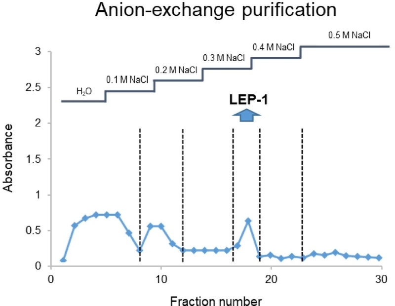
Purification scheme and chromatographic profiles of LEP-1.
Monosaccharide composition analysis – LEP-1 was hydrolyzed with 2 M TFA at 120°C for 12 h to release constituent monosaccharides. After hydrolysis, the reaction mixture was evaporated to dryness under a nitrogen stream. Residual TFA was removed by repeated dissolution in methanol followed by evaporation under reduced pressure. The dried residue was dissolved in deionized water and filtered through a 0.22 μm PTFE syringe filter prior to chromatographic analysis. Monosaccharide composition was determined using an ion chromatography system equipped with a Sugar-Pak™ I column (250 mm × 4.0 mm, 10 μm) and a refractive index (RI) detector. The column temperature was maintained at 60℃, and isocratic elution was performed with deionized water at a flow rate of 0.2 mL/min. Monosaccharides were identified and quantified by comparison with authentic standards, and relative molar percentages were calculated based on peak area normalization.
NMR spectroscopy – LEP-1 was dissolved in D₂O and NMR spectra were recorded on Bruker Avance III spectrometers (Waldbronn, Germany). One-dimensional 1H and 13C NMR spectra were acquired together with two-dimensional experiments, including 1H–1H COSY, TOCSY, ROESY, HSQC, and HSQC–TOCSY, for structural elucidation.
MALDI-TOF-MS analysis – MALDI-TOF-MS analysis of LEP-1 was performed using an Ultraflex II MALDI-TOF/TOF mass spectrometer (Bruker Daltonics, Germany) operated in positive-ion reflector mode. Spectra were acquired over an m/z range of 500–5000 using FlexControl software. The observed ion series was used to evaluate the oligomeric pattern of LEP-1.
Cell viability assay – The anti-proliferative activity of LEP-1 was evaluated using AGS human gastric adenocarcinoma cells. Cells were treated with various concentrations of LEP-1 for 24 h. After treatment, cell viability was assessed using the MTT assay according to standard procedures. The absorbance was measured at 570 nm using a microplate reader. Cell viability was expressed as a percentage relative to the untreated control group. IC₅₀ values were calculated using nonlinear regression analysis. All experiments were performed in triplicate.
Statistical analysis – All experiments were performed in triplicate. Data are presented as mean ± standard deviation (SD). Statistical significance was determined using one-way analysis of variance (ANOVA). Differences were considered statistically significant at p < 0.05.
Results and Discussion
Extraction and purification strategies critically influence the composition and purity of mushroom-derived polysaccharides.16,17 Hot-water extraction was selected to recover water-soluble glucan fractions under relatively mild conditions. Fractionation of the crude hot-water polysaccharide extract (HWPE) using a DEAE-Sephadex A-50 anion-exchange column yielded a major polysaccharide fraction, designated LEP-1 (Fig. 1).
Monosaccharide composition analysis was performed by comparison with authentic standards, and relative molar percentages were calculated based on peak area normalization (Fig. 2a). LEP-1 was found to consist exclusively of glucose, indicating that it is a homopolysaccharide. The absence of other monosaccharides suggests that LEP-1 belongs to the glucan class of mushroom polysaccharides.
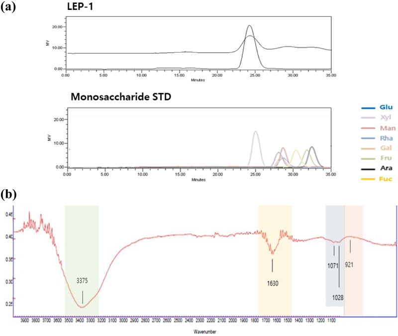
(a) Monosaccharide chromatogram and (b) FT-IR spectrum of LEP-1.
The FT-IR spectrum of LEP-1 exhibited characteristic features of polysaccharides (Fig. 2b). A broad absorption band at approximately 3375 cm⁻¹ was attributed to O–H stretching vibrations, reflecting the abundance of hydroxyl groups in the polysaccharide. The band at 1630 cm⁻¹ was attributed to absorbed water and/or O–H bending vibrations. Strong absorption bands at 1071 and 1028 cm⁻¹ were associated with C–O–C and C–O stretching vibrations of the polysaccharide backbone. In addition, an absorption band at approximately 921 cm⁻¹ was observed in the anomeric region. Together with the NMR data, these spectral features were consistent with a β-linked glucan structure. 18,19
The structural features of LEP-1 were further investigated using comprehensive 1D and 2D NMR analyses, including ¹H, ¹³C, HSQC-DEPT, COSY, TOCSY, and ROESY experiments (Table 1). The ¹H and ¹³C NMR spectra (Figs. 3a and 3b) exhibited resonances in the regions of δH 3.0–5.5 ppm and δC 60.0–105.0 ppm, respectively, characteristic of carbohydrate structures.19,20 Anomeric proton signals at δH 4.48–4.67 ppm showed correlations with carbon signals at δC 102.7–103.0 ppm in the HSQC-DEPT spectrum (Fig. 3c), indicating β-configured glucopyranosyl residues. No significant α-anomeric signals were observed, suggesting that LEP-1 is predominantly composed of β-linked glucose units.

1H and 13C NMR assignments for LEP-1 (δ, ppm in D2O).
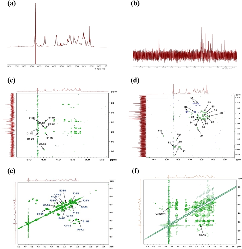
NMR spectrum of LEP-1. (a) 1H, (b) 13C NMR, (c) HSQC-DEPT, (d) HSQC-TOCSY, (e) COSY and (f) ROESY.
Downfield-shifted C-6 signals at δC 68.5 ppm, correlated with proton signals at δH 4.14 and 3.78 ppm, indicated substitution at O-6 and were assigned to →6)-β-D-Glcp-(1→ residues. A weaker substituted carbon signal at δC 84.3 ppm (δH 3.67 ppm) suggested the presence of →3)-β-D-Glcp-(1→ residues. The relatively stronger intensity of substituted C-6 signals compared with C-3 signals indicates that β-(1→6)-linked glucopyranosyl residues represent the dominant structural feature of LEP-1.
COSY and TOCSY spectra (Figs. 3d and 3e) enabled sequential assignment of proton resonances, while ROESY correlations (Fig. 3f) provided additional support for the proposed linkage pattern. Collectively, the monosaccharide composition and NMR data suggest that LEP-1 is a glucose-based β-glucan predominantly composed of →6)-β-D-glucopyranosyl residues with possible substitution at the O-3 position (Fig. 4). However, because methylation linkage analysis was not performed, the detailed linkage distribution and branching pattern of LEP-1 remain tentative and require further confirmation.
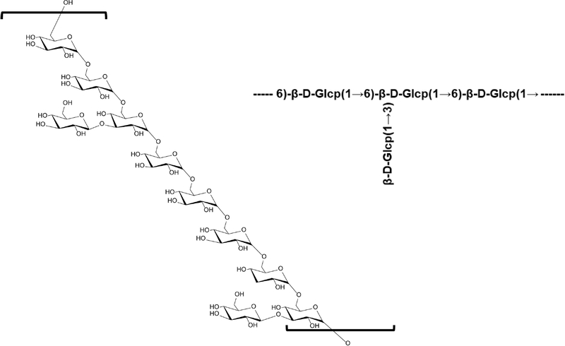
Tentative structural features of LEP-1 based on NMR analysis.
The molecular characteristics of LEP-1 were further investigated using NMR integration and MALDI-TOF-MS analysis (Fig. 5). The number-average degree of polymerization (DPn), estimated from the ratio of the total anomeric proton signals to the reducing-terminal anomeric proton signal, was approximately 22.
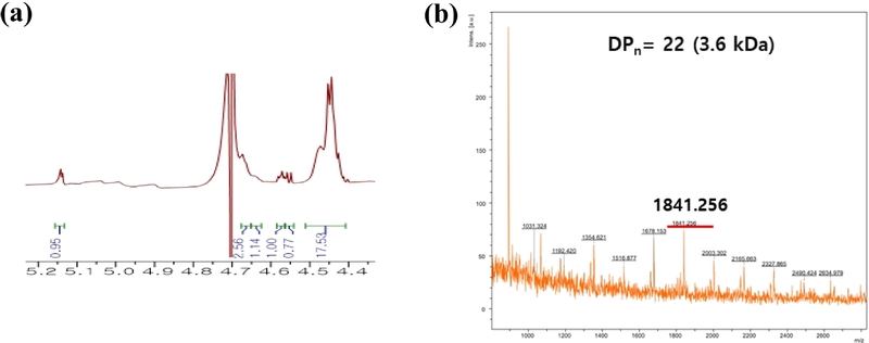
Molecular characterization of LEP-1. (a) 1H NMR and (b) MALDI–TOF–MS spectra of LEP-1.
MALDI-TOF-MS analysis in positive-ion mode revealed a series of sodium-adducted ions separated by approximately 162.1 Da, consistent with successive glucose units. The most intense ion at m/z 1841.256 corresponded approximately to an oligomer containing 11 glucose residues, which was lower than the NMR-derived DPn. This discrepancy may be attributed to preferential ionization and detection of lower-molecular-weight species in MALDI-TOF-MS and uncertainty in NMR integration arising from overlapping anomeric signals. Therefore, the MALDI-TOF-MS data were interpreted as supporting the presence of a glucan oligomer series rather than providing an absolute molecular-weight distribution of LEP-1. Further analysis using SEC/GPC with appropriate calibration or light-scattering detection would be required to accurately determine the molecular-weight distribution of LEP-1.
The antiproliferative activity of LEP-1 was evaluated against AGS human gastric adenocarcinoma cells using the MTT assay. LEP-1 reduced AGS cell viability in a concentration-dependent manner (Fig. 6). Cell viability was 86.0 ± 2.9%, 63.7 ± 5.0%, 55.0 ± 7.6%, and 44.7 ± 2.2% following treatment with 12.5, 25, 50, and 100 μg/mL LEP-1, respectively. The estimated IC₅₀ value was approximately 68.1 μg/mL. These results indicate that LEP-1 exhibits antiproliferative activity against AGS cells under the present experimental conditions. However, because the biological evaluation was limited to an MTT assay in a single cancer cell line, the observed effect should be regarded as preliminary antiproliferative activity.
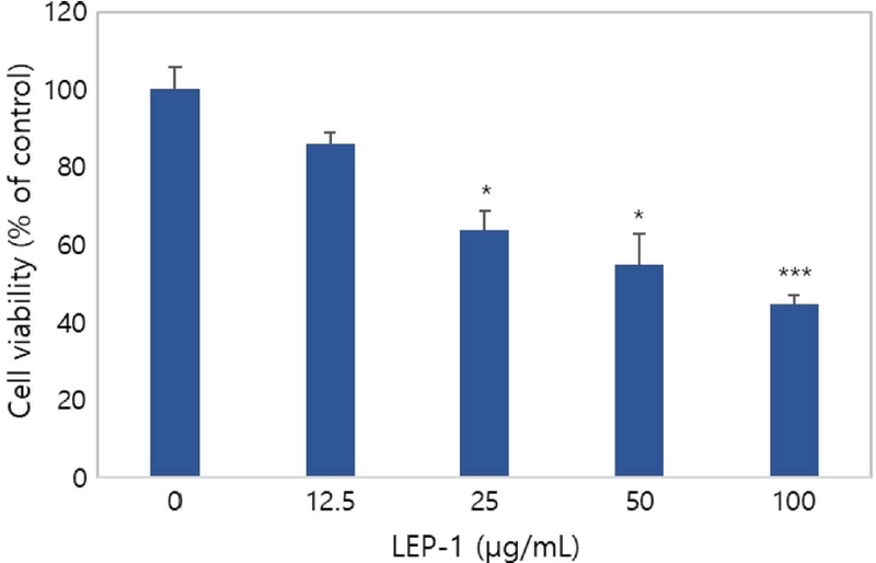
Effects of LEP-1 on the viability of AGS human gastric adenocarcinoma cells. Data are expressed as the mean ± SD of three independent experiments (n = 3). *p < 0.05 and ***p < 0.001 compared with the untreated control.
L. edodes has long been recognized as a rich source of bioactive polysaccharides, with lentinan being the most extensively studied example.11–13 Consequently, the structural characteristics and biological functions of shiitake polysaccharides have often been discussed primarily in the context of β-(1→3)-glucans containing β-(1→6)-linked branches. However, LEP-1 exhibited structural features distinct from the classical lentinan-type architecture, suggesting the presence of a previously underappreciated glucan population in L. edodes. This observation highlights a greater degree of glucan heterogeneity than is generally appreciated. Although additional studies will be required to fully elucidate the biological mechanism of LEP-1, the occurrence of LEP-1 further suggests that multiple glucan architectures coexist within the fruiting bodies of L. edodes and may contribute differently to their biological activities.
Acknowledgments
This work was supported by the Research grant (2022R1A2C1008081) of the National Research Foundation of Korea (NRF) funded by the Korean government (MSIT).
Conflicts of Interest
The authors declare that they have no conflicts of interest.
References
-
Nam, M.; Choi, J. Y.; Kim, M. S. Biomolecules 2021, 11, 1654.
[https://doi.org/10.3390/biom11111654]

-
Bugajewski, M.; Angerhoefer, N.; Pączek, L.; Kaleta, B. Int. J. Mol. Sci. 2025, 26, 3320.
[https://doi.org/10.3390/ijms26073320]
-
Xu, X.; Yan, H.; Tang, J.; Chen, J.; Zhang, X. Crit. Rev. Food Sci. Nutr. 2014, 54, 474–487.
[https://doi.org/10.1080/10408398.2011.587616]
-
Huang, X.; Nie, S. Food Funct. 2015, 6, 3205–3217.
[https://doi.org/10.1039/C5FO00678C]
-
Sheng, K.; Wang, C.; Chen, B.; Kang, M.; Wang, M.; Liu, K.; Wang, M. Food Chem. 2021, 358, 129883.
[https://doi.org/10.1016/j.foodchem.2021.129883]
-
Dai, Y.; Wang, L.; Chen, X.; Song, A.; He, L.; Wang, L.; Huang, D. Foods 2023, 12, 3289.
[https://doi.org/10.3390/foods12173289]
-
Zhao, Z. C.; Gu, F. T.; Li, J. H.; Zhu, Y. Y.; Huang, L. X.; Wu, J. Y. Int. J. Biol. Macromol. 2024, 280, 136082.
[https://doi.org/10.1016/j.ijbiomac.2024.136082]
-
Yu, Y.; Shen, M.; Song, Q.; Xie, J. Carbohydr. Polym. 2018, 183, 91–101.
[https://doi.org/10.1016/j.carbpol.2017.12.009]
-
Mostowy, R. J.; Holt, K. E. Trends Microbiol. 2018, 26, 1008–1021.
[https://doi.org/10.1016/j.tim.2018.06.006]
-
Zhao, J. Y.; Guo, J.; Ye, H. Y.; Luo, P. W.; Zhu, Q.; Xu, H.; Zhou, Y.; Wang, Y. J. Carbohydr. Polym. 2026, 371, 124463.
[https://doi.org/10.1016/j.carbpol.2025.124463]
-
Zhang, Y.; Li, S.; Wang, X.; Zhang, L.; Cheung, P. C. K. Food Hydrocoll. 2011, 25, 196–206.
[https://doi.org/10.1016/j.foodhyd.2010.02.001]
-
Zhang, L.; Li, X.; Xu, X.; Zeng, F. Carbohydr. Res. 2005, 340, 1515–1521.
[https://doi.org/10.1016/j.carres.2005.02.032]
-
Wang, Y.; Wang, H.; Chai, K.; Guo, S.; Zhai, Y.; Shi, R.; Zhang, F.; Huang, J.; Jin, Z.; Gao, Y.; Tao, X.; Yang, S.; Li, J.; Zhou, J.; Qiao, C.; Stalin, A.; Wu, J. Phytomedicine 2024, 123, 155242.
[https://doi.org/10.1016/j.phymed.2023.155242]
- Synytsya, A.; Novák, M. Ann. Transl. Med. 2014, 2, 17.
-
Wang, X.; Qu, Y.; Wang, Y.; Wang, X.; Xu, J.; Zhao, H.; Zheng, D.; Sun, L.; Tai, G.; Zhou, Y.; Cheng, H. Front. Immunol. 2022, 13, 859923.
[https://doi.org/10.3389/fimmu.2022.859923]
-
McCleary, B. V.; Draga, A. J. AOAC Int. 2016, 99, 364–373.
[https://doi.org/10.5740/jaoacint.15-0289]
-
Volman, J. J.; Ramakers, J. D.; Plat, J. Physiol. Behav. 2008, 94, 276–284.
[https://doi.org/10.1016/j.physbeh.2007.11.045]
- Kačuráková, M.; Capek, P.; Sasinková, V.; Wellner, N.; Ebringerová, A. Carbohydr. Polym. 2000, 43, 195–203.
- Wang, J.; Zhang, L.; Yu, Y.; Cheung, P. C. K. Int. J. Biol. Macromol. 2016, 92, 645–653.
-
Zhang, X.; Duan, Y.; Xue, J.; Chen, S.; Wang, H. Int. J. Biol. Macromol. 2025, 320, 145888.
[https://doi.org/10.1016/j.ijbiomac.2025.145888]