
Sclerotiorin-Type Azaphilones from Penicillium sp. with Their Anticancer Activities
Abstract
As part of ongoing efforts to discover fungal-derived antibacterial and anticancer natural products, a soil-derived fungal strain (Penicillium sp.) was identified as a hit based on its antibacterial activity against methicillin-resistant Staphylococcus aureus. Bioassay-guided fractionation of the active fungal extract led to the isolation of three sclerotiorin-type azaphilones, isochromophilone IX (1), isochromophilone VI (2), and sclerotioramine (3). The structures of 1–3 were identified by spectroscopic methods, including NMR spectroscopy and HR-ESI-Q-TOF-MS. Given the reported bioactivities of the azaphilone-class, the anticancer potential of the three isolated compounds was subsequently evaluated against five breast cancer cell lines representing four major molecular subtypes. As a result, compound 3 exhibited the most potent cytotoxicity, with IC50 values ranging from 26.08 to 47.06 μM, while compound 1 showed no activity (IC50 > 200 μM) against all cell lines tested, highlighting the importance of the isoquinoline N-substituent for cytotoxic activity.
Keywords:
Azaphilones, Soil-derived fungal isolate, Penicillium sp., Anticancer activity, Breast cancer cell linesIntroduction
Azaphilones represent a structurally diverse group of fungal polyketides, distinguished by a highly oxygenated pyranoquinone bicyclic core.1 Numerous ascomycetous and basidiomycetous fungal species are known producers of azaphilones.2 Sclerotiorin, a representative member of the azaphilone-class, was identified as a fungal metabolite of Penicillium sclerotiorum first3 and has since been isolated from various Penicillium species.3–9 Sclerotiorin-type azaphilones are characterized by a chlorinated isochromane core, including a conjugated ketone and a branched alkyl side chain.1 Various biological activities of sclerotiorin have previously been reported, including antibacterial,4 aldose reductase inhibitory,4 lipoxygenase production inhibitory,5 protein kinase G autophosphorylation inhibitory,10 and anticancer activities.11
Breast cancer is a heterogeneous group of tumors with considerable molecular and clinical diversity, resulting in differences in disease progression and therapeutic response. Breast cancers can be classified into Luminal A, Luminal B, Luminal A, Luminal B, human epidermal growth factor receptor 2 (HER2)-positive, and triple-negative subtypes-positive, and triple-negative subtypes based on hormone receptor and HER2 status.12,13 Hormone receptor-positive tumors are primarily treated with endocrine-based approaches, whereas HER2-positive tumors benefit from HER2-directed therapies.14 In contrast, the absence of estrogen receptor (ER), progesterone receptor (PR), and HER2 in triple negative breast cancers (TNBC) limits the applicability of these targeted treatments.15 In present study, the cytotoxic activities of compounds isolated from a soil-derived fungal strain identified as an antibacterial hit were evaluated using a panel of breast cancer cell lines representing Luminal A/B, HER2-positive, and TNBC subtypes to assess their anticancer potential across different breast cancer contexts. This approach was pursued because the antibacterial activities of the isolates have been already reported,6,16 whereas their cytotoxic effects against breast cancer cell lines remain poorly investigated despite having been evaluated against various other cancer cell lines (Table 1).

Comparison of cytotoxic activities of compounds 1–3 against various cancer cell lines from literature and the present study.
Experimental
General experimental procedures – Optical rotations were recorded on a JASCO P-2000 polarimeter (JASCO, Japan). The 1D and 2D NMR spectra were acquired on a Varian VNMRS 500 MHz FT-NMR instrument. HR-ESI-QTOF-MS was performed on an Agilent 1220 system coupled to Agilent 6540 UHD Accurate-Mass Q-TOF LC/MS system. Silica gel (230–400 mesh, Merck, Germany) and RP-18 (YMC gel ODS-A, 120 Å, S-150 μm, YMC Co., Japan) were used for column chromatography (CC). Thin layer chromatography (TLC) was performed using Kieselgel 60 F254 (silica gel, 0.25 mm layer thickness, Merck, Germany) and RP-18 F254S (Merck, Germany) plates detected on TLC under UV light (254 and 365 nm) followed by heating after spraying with 10% (v/v) sulfuric acid. Semipreparative high-performance liquid chromatography (HPLC) was conducted on a Waters system using a Waters 1525 binary pump with a Waters 2996 photodiode array detector, using a YMC-Triart C18 column (5 μm, 250 mm × 10.0 mm i.d.).
Fungal materials – The fungal strain DS2-11 was isolated from the soil sample collected from a medicinal herb garden near Duksung Women’s University (Seoul, Republic of Korea). The fungal strain was identified as Penicillium sp. (GenBank Accession No. PQ130407, P. sclerotiorum with 99.1% identity) based on the ribosomal internal transcribed spacer (ITS) region (Biofact Inc.).17
Extraction and isolation – Fungal cultivation, extraction, and initial fractionation were performed as previously reported.17 Briefly, the fungal culture of Penicillium sp. [DS2-11] was homogenized and extracted three times with an equal volume of ethyl acetate (EtOAc). The resulting EtOAc-soluble extract (9 g) was fractionated by silica gel vacuum liquid chromatography (VLC), eluting stepwise with dichloromethane (DCM), DCM/methanol (MeOH) (10:1), and MeOH to afford three fractions. The DCM/MeOH (10:1) fraction was further separated by HP20SS VLC using a stepwise gradient of MeOH/H2O (3:7, 1:1, 7:3, 9:1, and 1:0) followed by DCM/MeOH (1:1), yielding six fractions. A part (500 mg) of the MeOH/H2O (9:1) fraction (1.88 g) from HP20SS VLC was chromatographed over RP-18, using a MeOH/H2O (7:3 → 9:1) step gradient solvent mixtures, to yield 13 subfractions (H1-H13). Fraction H2 (17.2 mg) was further purified using semipreparative HPLC [acetonitrile (MeCN)/H2O containing 0.1% formic acid 55:45, 2 mL/min] to yield 1 (2.3 mg, tR 23.5 min). Compound 2 (3.2 mg) was separated from fraction H4 (27.3 mg) by semipreparative HPLC with MeCN/H2O containing 0.1% formic acid (55:45, 2 mL/min, tR 24.5 min). A part (10.0 mg) of fraction H6 (360.1 mg) was subjected to RP-18 semipreparative HPLC (MeCN/H2O containing 0.1% formic acid 55:45, 2 mL/min), to afford compound 3 (4.1 mg, tR 25.7 min).
Isochromophilone IX (1) – Red amorphous solid; +43.49 (c 0.10, MeOH); UV (HPLC) λmax 490, 362, 238 nm; 1H and 13C NMR data, Table 2; HR-ESI-QTOF-MS m/z 476.1855 [M + H]+ (calcd for C25H31ClNO6, 476.1834).7,18
1H (500 MHz) and 13C (125 MHz) NMR spectroscopic data of isochromophilone IX (1), isochromophilone VI (2) and sclerotioramine (3) in CD3OD.
Isochromophilone VI (2) – Red amorphous solid; +12.86 (c 0.10, MeOH); UV (HPLC) λmax 490, 363, 238 nm; 1H and 13C NMR data, Table 2; HR-ESI-QTOF-MS m/z 434.1744 [M + H]+ (calcd for C23H29ClNO5, 434.1729).7,18
Sclerotioramine (3) – Red amorphous solid; +28.7 (c 0.10, MeOH); UV (HPLC) λmax 497, 348, 232 nm; 1H and 13C NMR data, Table 2; HR-ESI-QTOF-MS m/z 390.1485 [M + H]+ (calcd for C21H25ClNO4, 390.1467).9,18
Quantum mechanics-driven 1H iterative full spin analysis (QM-HiFSA) – HiFSA calculations were carried out using CT (Cosmic Truth), a web-based software developed by NMR Solutions (Kuopio, Finland).19 Prior to the calculations, the 1H NMR data in JCAMP-DX (.jdx) format were prepared using MestReNova 14.0.0 software (Mestrelab Research SL, Santiago de Compostela, Spain), with preprocessing including zero-filling to 256k, referencing to the residual solvent signal at δH 3.3100 ppm, fifth-order polynomial baseline correction, and Lorentzian−Gaussian apodization (line broadening = − 0.1, Gaussian factor = 0.2). In addition, for QM-based prediction in CT, 3D MOL files of the chemical structures were constructed using MM2 energy minimization in Chem 3D Ultra software (PerkinElmer Informatics, Waltham, MA, USA).20
Cell culture – Human breast cancer cell lines, i.e., MCF7, T47D, BT474, SKBR3, and MDA-MB-231, were obtained from the American Type Culture Collection (ATCC). MCF7, BT474, and MDA-MB-231 cells were maintained in Dulbecco’s modified Eagle’s medium supplemented with 10% fetal bovine serum (FBS). T47D and SKBR3 cells were maintained in RPMI-1640 medium supplemented with 10% FBS. The cells were grown in an incubator with 5% CO2/95% air at 37°C and routinely certified free of mycoplasma contamination using the MycoAlert Mycoplasma Detection Kit (Lonza).
Cell viability test – Cells were seeded in 96-well plates and treated with increasing concentrations of the isolated compounds (1–3) or doxorubicin (positive control) for 24 h. Following treatment, 3-(4,5-dimethylthiazol-2-yl)-2,5-diphenyltetrazolium bromide (MTT) solution was added, and cells were incubated at 37°C for formazan formation. The formazan crystals were dissolved in dimethyl sulfoxide, and absorbance was measured at 570 nm with 750 nm as the reference wavelength. Cell viability was calculated from the background-corrected absorbance (OD570−OD750) and expressed as a percentage of the vehicle-treated control. IC50 values were determined by four-parameter nonlinear regression using GraphPad Prism (version 10.6.1). Data are presented as mean ± SD (n = 3).
Results and Discussion
In our continued search for fungal bioactive natural products,17 60 soil-derived fungal isolates were evaluated for antibacterial activity against methicillin-resistant Staphylococcus aureus (MRSA). Among them, one fungal isolate was selected as a hit and identified as Penicillium sp. (P. sclerotiorum based on 99.1% ITS sequence identity). Subsequent bioassay-guided fractionation of its active extract yielded three metabolites (Fig. 1) as red amorphous solids, all of which showed similar UV absorption profiles with λmax near 240, 360, and 490 nm. The isolated compounds 1–3 were identified as the sclerotiorin-type azaphilones isochromophilone IX, isochromophilone VI, and sclerotioramine, respectively, based on interpretation of 1D and 2D NMR data and comparison with literature values.7,9 This study reports the 1H and 13C NMR data acquired in CD3OD (Table 2), rather than in CDCl3 as previously reported for these compounds, since they showed low solubility in CDCl3 in the present study. Therefore, analysis of 2D NMR data (1H-1H COSY, 1H-13C HSQC, and 1H-13C HMBC NMR) was performed to provide accurate assignments. In addition, a more detailed and accurate description of previously reported multiplets was achieved by using QM-HiFSA calculations. HR-ESI-QTOF-MS data obtained for 1–3 further supported the presence of nitrogen rather than the pyran oxygen found in sclerotiorin (Fig. 2). Furthermore, the characteristic chlorine isotopic pattern was clearly observed, with [M + H]+ and [M + H + 2]+ peaks in an approximate ratio of 3:1.18 Compound 1 showed [M + H]+ (m/z 476.1855) and [M + H + 2]+ (m/z 478.1834) ion peaks. Similarly, compound 2 produced [M + H]+ and [M + H + 2]+ ions at m/z 434.1744 and 436.1722, respectively. Compound 3 displayed a molecular ion peak at m/z 390.1485 [M + H]+ with 3:1 chlorine isotope peak at m/z 392.1461 [M + H + 2]+.

Chemical structures of isochromophilone IX (1), isochromophilone VI (2), and sclerotioramine (3).
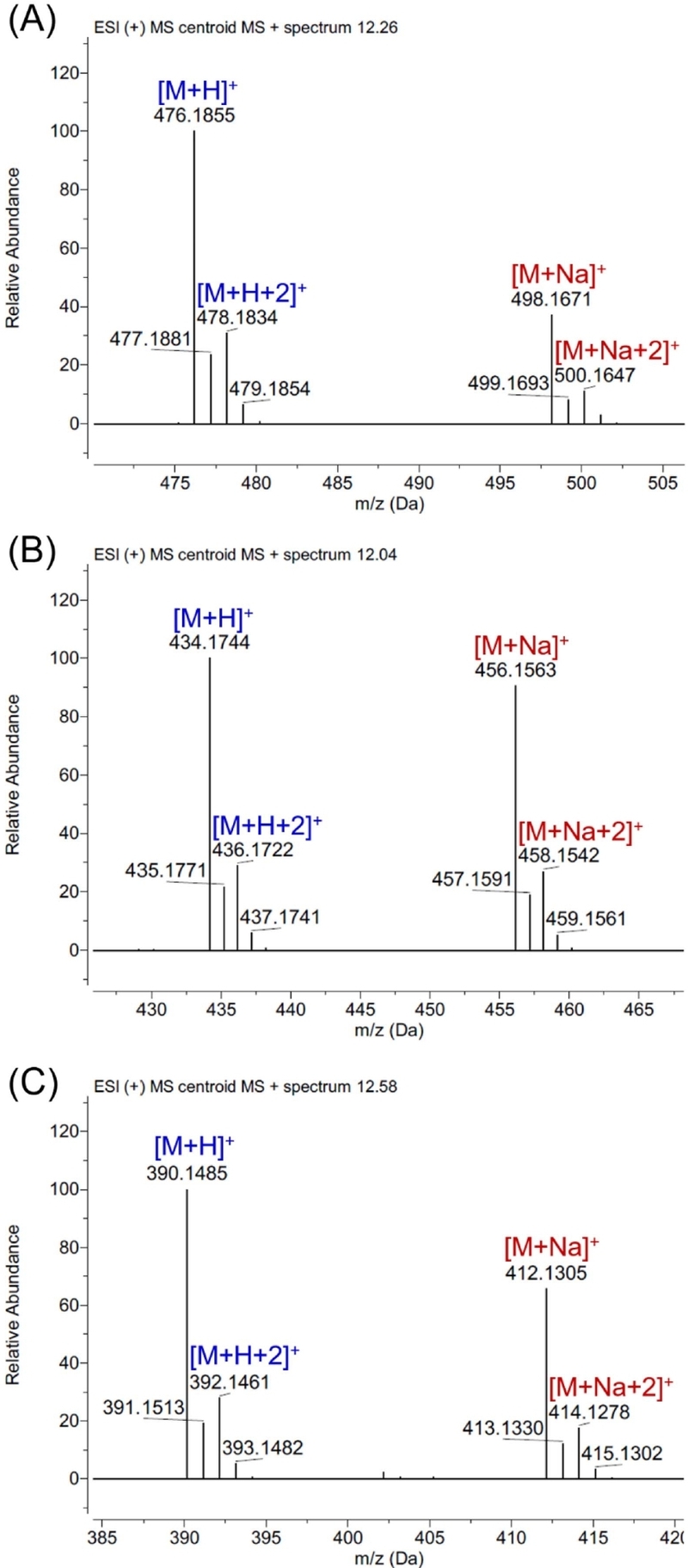
Expanded HR-ESI-Q-TOF-MS spectra of (A) isochromophilone IX (1), (B) isochromophilone VI (2), and (C) sclerotioramine (3).
As the three fungal metabolites share many structural features (Fig. 1), 1H and 13C NMR data of 1–3 (Table 2) were also closely comparable. Accordingly, the common structural features can be described using the data of isochromophilone IX (1). The 1H NMR spectrum of 1 showed two olefinic protons signals at δH 8.2205 (1H, s, H-1) and 7.2126 (1H, s, H-4), with a methyl signal at δH 1.5084 (3H, s, H3-18) located on isoquinoline ring, and an additional methyl signal at δH 2.1177 (3H, s, H3-20) assigned to an acetate group attached to the ring. In the 13C NMR spectrum, two conjugated ketone carbonyl carbons were observed at δC 185.4 (C-6) and 195.0 (C-8), characteristic of the azaphilone class. The additional three olefinic methine protons at δH 6.6321 (1H, d, J = 15.37 Hz, H-9), 7.1558, (1H, d, J = 15.37 Hz, H-10) and 5.7949 (1H, d, J = 9.72 Hz, H-12) were attributed to the branched alkyl side chain. Two nonequivalent methylene protons were observed at δH 1.4753 (1H, dqd J = -13.52, 7.32, 5.37 Hz, H-14a), 1.3654 (1H, ddq J = -13.52, 8.35, 7.51 Hz, H-14b), along with three methyl signals at δH 0.9037 (3H, dd, J = 7.51, 7.32 Hz, H3-15), 1.0413 (3H, d, J = 6.62 Hz, H3-16), 1.9682 (3H, s, H3-17). All these resonances indicated the presence of the 3,5-dimethyl-1,3-heptadien-1-yl group.7,9,18
While sharing the common structural features described above, compounds 1–3 differ only in the N-substituent at the isoquinoline nitrogen. The 1H NMR spectrum of 1 showed resonances attributable to a 3-carboxypropyl group at δH 4.2134 (1H, ddd, J = -13.65, 6.96, 6.87 Hz, H-1′a), 4.1759 (1H, ddd, J = -13.65, 7.64, 7.02 Hz, H-1′b), 2.0552 (1H, ddddd, J = -14.00, 8.16, 7.64, 6.96, 6.25 Hz, H-2′a), 2.0389 (1H, ddddd, J = -14.00, 8.05, 7.02, 6.87, 6.25 Hz, H-2′b) and 2.4101 (1H, ddd, J = -16.39, 8.16, 6.25 Hz, H-3′a), 2.4057 (1H, ddd, J = -16.39, 8.05, 6.25 Hz H-3′b).7 Compound 2 contained a 2-hydroxyethyl group, with resonances observed at δH 4.2573 (1H, ddd, J = -14.77, 5.48, 4.25 Hz, H-1′a), 4.2301 (1H, ddd, J = -14.77, 5.94, 4.06 Hz, H-1′b) and 3.8402 (1H, ddd, J = -9.30, 4.25, 4.06 Hz, H-2′a), 3.8380 (1H, ddd, J = -9.30, 5.94, 5.48 Hz, H-2′b). In compound 3, the isoquinoline nitrogen remained unsubstituted as an NH group.
These sclerotiorin-type azaphilones (1–3) were initially isolated from bioassay-guided fractionation for antibacterial activity against MRSA in the present study. However, as their antibacterial activity against MRSA had already been reported,6,16 based on literature, our attention turned to the anticancer potential of these metabolites. In the development of anticancer agents, selective activity towards specific cancer cells or subtypes is considered an important factor. Therefore, it is valuable to explore other cancer cell lines that have not yet been evaluated in terms of selective cytotoxicity.21 To provide context for previously reported cytotoxicity of 1–3, cancer cell lines tested were reviewed and summarized in Table 1, which included a diverse panel of cancer cell lines derived from breast (MCF-7, MDA-MB-435, MDA-MB-231),11,17,22 cervical (HeLa),8 promyelocytic leukemia (HL-60),8 liver (HepG2),23 papilloma (KB),11 and lung (NCI-H187, A549).11,22 Among them, the cell line MDA-MB-435 was historically classified as a breast cancer cell line but was subsequently demonstrated to be of melanoma origin.24 Our literature review indicated that the cytotoxicity of sclerotiorin-type azaphilones against breast cancer cell lines remains largely unexplored, with only sclerotioramine (3) previously evaluated against the triple-negative breast cancer cell line MDA-MB-231.17 Accordingly, in this study, the cytotoxic activities of compounds 1–3 were evaluated against five breast cancer cell lines (Fig. 3): MCF7, T47D and BT474 (Luminal A/B), SKBR3 (HER2-positive), and MDA-MB-231 (triple-negative breast cancer, TNBC). As each subtype relies on distinct signaling pathways and therapeutic approaches, evaluating compounds across multiple breast cancer subtypes than a single cell line enabled the identification of both subtype specific and broad-spectrum activities.6,12–16
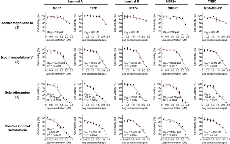
Cytotoxicity of compounds 1–3 against Luminal A subtype cell lines MCF-7 and T47D, Luminal B subtype cell line BT474, HER2-positive subtype cell line SKBR3, and TNBC subtype cell line MDA-MB-231.
Compound 1 displayed no cytotoxicity (IC50 > 200 μM) against any of the tested cell lines, whereas compounds 2 and 3 showed varying degrees of activity, with 3 consistently outperforming 2 across all subtypes tested (Table 1 and Fig. 3). Three isolates share the same sclerotiorin-type azaphilone scaffold but differ in N-substituents. The results showed that compound 3, which contains the unsubstituted NH group, exhibited greater cytotoxicity against breast cancer cell lines than compounds 1 and 2, which contain 3-carboxypropyl and 2-hydroxyethyl groups, respectively. This suggested that the N-substituent may contribute to cytotoxic activity, although additional studies would be required to establish a definitive structure-activity relationship. In addition, compounds 2 and 3 were relatively more active against the Luminal A/B cancer cell lines (MCF-7 and T47D) than against the other subtypes tested.
Compound 2 exhibited IC50 values of 59.43–81.56 μM in the breast cancer cell lines examined (Table 1 and Fig. 3), and to our knowledge, it has not previously been evaluated for cytotoxicity against authenticated breast cancer cell lines. In the previous studies, compound 2 displayed notable potency against human cervix cancer cell line HeLa (IC50 = 21.4 μM) and human promyelocytic leukemia cell line HL-60 (IC50 = 17.7 μM).8 These results may be explained by the effect of the longer 48 h treatment period used in the previous studies; however, they may also suggest cell type-dependent cytotoxic activity of 2. Sclerotioramine (3) has been reported to have potent cytotoxic effects against human liver cancer cell line HepG2 (IC50 = 15.4 μM),23 human papilloma cancer cell line KB (IC50 = 4.6 μM),11 human lung cancer cell line NCI-H187 (IC50 = 11.6 μM),11 which was consistent with its being the most potent of the three compounds in our breast cancer cell line evaluation (IC50 values of 26.08–47.06 μM). Compound 3 showed an IC50 value of 34.46 μM against the triple-negative breast cancer cell line MDA-MB-231 in this study, which was higher than the previously reported value of 21.8 μM in the same cell line after 48 h treatment,17 while stronger cytotoxicity was observed against MCF-7 cells (IC50 = 26.08 μM) than was previously reported (IC50 = 34.7 μM), possibly due to differences in experimental conditions.
In the present study, three sclerotiorin-type azaphilones (1–3) were evaluated for their cytotoxicity against a panel of breast cancer cell lines. This study extended previous evaluations of the anticancer activity of the sclerotiorin-type azaphilones; however, no selective cytotoxicity toward the breast cancer cell lines was observed. In addition, previous reports on the cytotoxic activity of isochromophilone IX (1) have been limited, and it showed no notable cytotoxic activity against any of the breast cancer cell lines tested in this study. Thus, the present study provided an expanded cytotoxicity profile for 1, while comparative evaluation of the three compounds indicated that the type of N-substituent may play a critical role in the cytotoxic activity of sclerotiorin-type azaphilones.
Acknowledgments
This work was supported by the National Research Foundation of Korea (NRF) grant funded by the Korean government (MSIT) in 2026 (Grant No. RS-2026-25601128, Project Title: Discovery of New Natural Immune-Modulating Agents for Cancer Immunotherapy). And following are results of a study on the "Gyeongsangnam-do Regional Innovation System & Education (RISE)" Project, supported by the Ministry of Education and Gyeongsangnam-do (2026-RISE-16-008-0016). This project was also supported by a grant from Chonnam National University (2024-1948-03).
Conflicts of Interest
The authors declare that they have no conflicts of interest.
References
-
Gao, J.-M.; Yang, S.-X.; Qin, J.-C. Chem. Rev. 2013, 113, 4755–4811.
[https://doi.org/10.1021/cr300402y]

-
Gill, M.; Steglich, W. Pigments of Fungi (Macromycetes); Springer Vienna, 1987; 1–297.
[https://doi.org/10.1007/978-3-7091-6971-1_1]
-
Curtin, T. P.; Reilly, J. Biochem. J. 1940, 34, 1411–1421.
[https://doi.org/10.1042/bj0341605]
-
Chidananda, C.; Rao, L. J. M.; Sattur, A. P. Biotechnol. Lett. 2006, 28, 1633–1636.
[https://doi.org/10.1007/s10529-006-9133-4]
-
Chidananda, C.; Sattur, A. P. J. Agric. Food Chem. 2007, 55, 2879–2883.
[https://doi.org/10.1021/jf062032x]
-
Liu, Z.; Qiu, P.; Liu, H.; Li, J.; Shao, C.; Yan, T.; Cao, W.; She, Z. Bioorg. Chem. 2019, 88, 102973.
[https://doi.org/10.1016/j.bioorg.2019.102973]
-
Michael, A. P.; Grace, E. J.; Kotiw, M.; Barrow, R. A. Aust. J. Chem. 2003, 56, 13–15.
[https://doi.org/10.1071/CH02021]
-
Son, S.; Ko, S.-K.; Kim, J. W.; Lee, J. K.; Jang, M.; Ryoo, I.-J.; Hwang, G. J.; Kwon, M. C.; Shin, K.-S.; Futamura, Y.; et al. Phytochemistry 2016, 122, 154–164.
[https://doi.org/10.1016/j.phytochem.2015.12.008]
-
Wang, X.; Sena Filho, J. G.; Hoover, A. R.; King, J. B.; Ellis, T. K.; Powell, D. R.; Cichewicz, R. H. J. Nat. Prod. 2010, 73, 942–948.
[https://doi.org/10.1021/np100142h]
-
Chen, D.; Ma, S.; He, L.; Yuan, P.; She, Z.; Lu, Y. Tuberculosis 2017, 103, 37–43.
[https://doi.org/10.1016/j.tube.2017.01.001]
-
Hemtasin, C.; Kanokmedhakul, S.; Moosophon, P.; Soytong, K.; Kanokmedhakul, K. Phytochem. Lett. 2016, 16, 56–60.
[https://doi.org/10.1016/j.phytol.2016.03.004]
-
Esposito, A.; Criscitiello, C.; Omodeo Salè, E.; Curigliano, G. Expert Rev. Clin. Pharmacol. 2014, 7, 605–611.
[https://doi.org/10.1586/17512433.2014.945429]
-
Sørlie, T.; Perou, C. M.; Tibshirani, R.; Aas, T.; Geisler, S.; Johnsen, H.; Hastie, T.; Eisen, M. B.; van de Rijn, M.; Jeffrey, S. S.; et al. Proc. Natl. Acad. Sci. U. S. A. 2001, 98, 10869–10874.
[https://doi.org/10.1073/pnas.191367098]
-
Zardavas, D.; Irrthum, A.; Swanton, C.; Piccart, M. Nat. Rev. Clin. Oncol. 2015, 12, 381–394.
[https://doi.org/10.1038/nrclinonc.2015.73]
-
Dent, R.; Trudeau, M.; Pritchard, K. I.; Hanna, W. M.; Kahn, H. K.; Sawka, C. A.; Lickley, L. A.; Rawlinson, E.; Sun, P.; Narod, S. A. Clin. Cancer Res. 2007, 13, 4429–4434.
[https://doi.org/10.1158/1078-0432.CCR-06-3045]
-
Shen, S. J.; Feng, Z. Y.; Jiang, S. J.; Liu, L.; Fu, S. J.; Chen, W. H.; Sun, Q. Y.; Chen, J. J. Chem. Biodivers. 2023, 20, e202201180.
[https://doi.org/10.1002/cbdv.202201180]
-
Kim, E.-S.; Lee, J. W. Nat. Prod. Sci. 2025, 31, 32–42.
[https://doi.org/10.1039/D5NP00027K]
-
Lu, T.; Liu, Y.; Zhou, L.; Liao, Q.; Nie, Y.; Wang, X.; Lei, X.; Hong, P.; Feng, Y.; Hu, X.; et al. Front. Microbiol. 2023, 14, 1144323.
[https://doi.org/10.3389/fmicb.2023.1144328]
-
Shao, Y.; Molnar, L. F.; Jung, Y.; Kussmann, J.; Ochsenfeld, C.; Brown, S. T.; Gilbert, A. T. B.; Slipchenko, L. V.; Levchenko, S. V.; O’Neill, D. P.; et al. Phys. Chem. Chem. Phys. 2006, 8, 3172–3191.
[https://doi.org/10.1039/B517914A]
-
Kil, Y.-S.; Park, J.; Jeong, B.-S.; Thapa, P.; Ok, Y. J.; Choi, H.; Jeong, J.-H.; Nam, J.-W. ACS Omega 2025, 10, 13185–13192.
[https://doi.org/10.1021/acsomega.4c10696]
-
Wilding, J. L.; Bodmer, W. F. Cancer Res. 2014, 74, 2377–2384.
[https://doi.org/10.1158/0008-5472.CAN-13-2971]
-
Chen, T.; Huang, Y.; Hong, J.; Wei, X.; Zeng, F.; Li, J.; Ye, G.; Yuan, J.; Long, Y. Mar. Drugs 2020, 19, 12.
[https://doi.org/10.3390/md19010012]
-
Yuan, W.-H.; Wei, Z.-W.; Dai, P.; Wu, H.; Zhao, Y.-X.; Zhang, M.- M.; Jiang, N.; Zheng, W.-F. Chem. Biodivers. 2014, 11, 1078–1087.
[https://doi.org/10.1002/cbdv.201300349]
-
Korch, C.; Hall, E. M.; Dirks, W. G.; Ewing, M.; Faries, M.; Varella-Garcia, M.; Robinson, S.; Storts, D.; Turner, J. A.; Wang, Y.; et al. Int. J. Cancer 2018, 142, 561–572.
[https://doi.org/10.1002/ijc.31067]