
Antiproliferative Effect of γ-Mangostin in Gastric Cancer Cells is Mediated by c-Met Signaling Pathway
Abstract
Encoding the receptor for hepatocyte growth factor (HGF), the proto-oncogene c-Met represents one of the tyrosine kinases most commonly found to be dysregulated across human cancers, and has consequently emerged as a promising target for anticancer drug discovery. γ-Mangostin, one of the major bioactive constituents of mangosteen (Garcinia mangostana L.), is known to regulate a range of cancer-related signaling pathways. MKN45 gastric cancer cell line, harboring c-Met amplification, were treated with γ-mangostin and evaluated by western blot, cell viability and apoptosis assays. We further confirmed that γ-mangostin treatment modulates the expression of c-Myc, cyclin D1, and p21, and suppresses the activity of pro-survival proteins, specifically Bcl-2 and Bcl-xL, through downregulation of c-Met. γ-Mangostin suppressed cell proliferation in MKN45 with an IC50 values 32.6 μM. Relative to untreated controls, γ-mangostin exposure led to elevated p21 Waf1/Cip1 levels and reduced expression of c-Myc, CD44, and cyclin D1. In addition, γ-mangostin suppressed the constitutive phosphorylation of c-Met, as well as that of its downstream signaling proteins ERK1/2 and STAT3. Anti-oncogenic and apoptotic effects (c-Myc, p21 Waf1/Cip1 and Caspase-3) by γ–mangostin treatment are mediated by c-Met inactivation. In conclusion, these observations indicate the potential of γ–mangostin as an effective antitumor agent against gastric adenocarcinoma. In addition, these results suggest that the compound's antitumor activity is mediated, at least in part, through suppression of c-Met signaling.
Keywords:
γ-Mangostin, MKN45, c-Met, Garcinia mangostanaIntroduction
A wide range of tropical plants exhibit intriguing biological properties that hold potential for therapeutic use. One such example is Garcinia mangostana L. (Clusiaceae), or mangosteen, which is extensively utilized as a botanical dietary supplement globally, typically marketed and consumed as a fruit juice.1,2 As one of the principal bioactive compounds found in mangosteen fruit, γ-mangostin has been shown to possess diverse biological properties, such as anti-inflammatory, antibacterial, and anticancer effects.3–7
c-Met signaling is known to be associated with several human cancers and a refined understanding of its molecular basis has promoted the development of a various antagonists with clinical potential.8 As the HGF (hepatocyte growth factor) receptor located on the cell surface, c-Met tyrosine kinase mediates intracellular signaling associated with cell proliferation, suppression of apoptosis, and mitogenesis.9 Well characterized downstream signaling pathways that are activated by c-Met kinase include the ERK and STAT3 pathways, resulting in proliferation.10 Furthermore, c-Met overexpression has been implicated in the pathogenesis of diverse human cancers, reflecting its broad relevance as a therapeutic target across multiple tumor types.11,12 All of these emphasize c-Met as an attractive target for cancer therapy.13
Effects of xanthone components extracted from mangosteen in cancer have been studied recently as described by Shan et al.14 In particular, the anticancer effects of γ-mangostin have been extensively investigated in both in vitro and in vivo studies4. Antiproliferative effects of γ-mangostin were also reported in DLD-1 colon cancer cell line.15 In this study, the effect of γ-mangostin on the gastric cancer cells was evaluated. γ-Mangostin showed potent cytotoxic activity, and expression pattern change of apoptotic protein and cell cycle protein by γ-mangostin was consistent with this finding. Further mechanistic study revealed that cytotoxic effects of γ-mangostin were mediated by c-Met signaling pathway. The present study will explain the biological effects of mangosteen and extend the use of mangosteen as dietary supplement.
Experimental
General experimental procedures – Dulbecco’s modified Eagle’s medium (DMEM), fetal bovine serum (FBS), penicillin, and streptomycin were supplied by HyClone (Logan, UT, USA). Bovine serum albumin (BSA) and dimethyl sulfoxide (DMSO) were obtained from Sigma-Aldrich (St. Louis, MO, USA). Primary antibodies recognizing phosphorylated c-Met, total c-Met, cyclin D1, c-Myc, p21 Waf1/Cip1, Bcl-2, Bcl-xL, and β-actin were sourced from Cell Signaling Technology, Inc. (Danvers, MA, USA). Oligonucleotide primers were synthesized by Bioneer Corp. (Daejeon, Korea). α-Mangostin and γ-mangostin were isolated from 700 g of Garcinia mangostana pericarp according to a previously described method, and their identities were established by comparing the acquired 1H and 13C NMR spectroscopic data with literature values.5
Cell culture and viability assay – MKN45 gastric carcinoma cells of human origin were propagated in Dulbecco’s modified Eagle’s medium containing 10% fetal bovine serum. For gene-silencing experiments, the cells were introduced with either c-Met-specific small interfering RNA or a nonspecific control oligonucleotide using Lipofectamine 2000 (Life Technologies, Carlsbad, CA, USA), following the instructions supplied with the transfection reagent. Both siRNA preparations were obtained from Santa Cruz Biotechnology, Inc. (Santa Cruz, CA, USA). The effects of the test compounds on cell survival were assessed with a WST-1-based colorimetric method (Roche, Laval, QC, Canada). Cells were distributed into 96-well culture plates (Nunc, Denmark) at 1 × 10⁵ cells/mL and allowed to undergo treatment with a series of compound concentrations for the designated incubation periods. Wells receiving the corresponding amount of DMSO without test compound were used as vehicle controls. After completion of the reaction, optical density was recorded on a microplate spectrophotometer manufactured by Molecular Devices Corporation (Sunnyvale, CA, USA). The concentration required to reduce cell viability by 50% was derived from the concentration–response relationship using regression analysis.
Western blot analysis – Cells were harvested and lysed, and the protein content of each lysate was measured using the Bio-Rad protein assay reagent (Bio-Rad, Richmond, CA, USA). Each lysate containing 20 μg of protein was combined with an equal volume of 2× loading buffer composed of 20% glycerol, 4% SDS, 10% 2-mercaptoethanol, 0.05% bromophenol blue, and 1.25 M Tris-HCl (pH 6.8). Proteins were resolved on 8% or 15% SDS-polyacrylamide gels by electrophoresis at 150 V for 90 min and transferred to polyvinylidene difluoride membranes using a Bio-Rad semi-dry transfer apparatus. The membranes were incubated overnight with the relevant primary antibodies, diluted between 1:500 and 1:1,000 in Tris-buffered saline supplemented with 0.1% Tween 20. After three washes with the same buffer, HRP-conjugated secondary IgG antibodies, diluted from 1:2,000 to 1:20,000, were applied for 1 h. The membranes were then washed three additional times, and antibody-bound proteins were detected using a chemiluminescent substrate from Amersham Biosciences (Piscataway, NJ, USA).
Quantitative real-time RT-PCR – Total RNA was prepared from the cells with TRIzol reagent (Life Technologies, CA, USA) according to the supplier’s instructions. For cDNA synthesis, 1 μg of RNA was incubated with 200 U of reverse transcriptase (Promega, Madison, WI, USA) and 500 ng of an oligo(dT) primer. The reverse-transcription mixture contained 50 mM Tris-HCl at pH 8.3, 75 mM KCl, 3 mM MgCl2, 10 mM DTT, and 1 mM dNTPs. The reaction was allowed to proceed at 42°C for 60 min, followed by enzyme inactivation at 70°C for 15 min. Quantitative PCR was performed in a final volume of 10 μL using 1 μL of the synthesized cDNA, 9 μL of a reaction mixture containing iQ SYBR Green Supermix (Bio-Rad), and 5 pmol each of the forward and reverse primers. Amplification was carried out on a CFX384 Real-Time PCR Detection System (Bio-Rad). The thermal program consisted of 3 min at 95°C, followed by 40 amplification cycles comprising 10 s at 95°C and 30 s at 55°C. SYBR Green fluorescence was monitored during each cycle, and the formation of a single amplification product was confirmed by melt-curve analysis. Threshold cycle values were obtained using CFX Manager software (Bio-Rad). GAPDH was used as the internal reference gene, and relative transcript levels were determined by the 2−ΔΔCt method. The following specific primer sets were used (5′ to 3′): c-Myc: TTTCGGGTAGTGGAAAACCA (forward), CACCGAGTCGTAGTCGAGGT (reverse); GAPDH: GAAGGTGAAGGTCGGAGTCA (forward), AATGAAGGGGTCATTGATGG (reverse); p21 Waf1/Cip1: TGCGTTCACAGGTGTTTCTG (forward), GCCATTAGCGCATCACAGT (reverse); Cyclin D1: GCGGAGGAGAACAAACAGAT (forward), GGCGGATTGGAAATGAACT (reverse). Gene-specific primers were custom-synthesized by Bioneer (Daejeon, Korea).
Immunofluorescence – MKN45 cells were cultured in Permanox chamber slides (Nunc, Rochester, NY, USA) and fixed with ethanol at 4°C for 30 min. After rinsing with phosphate-buffered saline (PBS), nonspecific binding sites were blocked with 3% bovine serum albumin in PBS for 30 min. The cells were then exposed to a rabbit monoclonal antibody against p21 Waf1/Cip1 at a dilution of 1:500 and maintained at 4°C overnight. Following several PBS washes, an Alexa Fluor 488-labeled secondary antibody obtained from Santa Cruz Biotechnology was applied for 2 h at room temperature. The stained specimens were washed again with PBS and mounted in ProLong Gold Antifade reagent containing 4′,6-diamidino-2-phenylindole (DAPI; Thermo Scientific, USA) for nuclear visualization. Fluorescence images were collected using an LSM510 confocal laser-scanning microscope (Carl Zeiss, Germany).
In vitro kinase assay – The ability of the test compounds to inhibit c-Met enzymatic activity was examined using a recombinant kinase assay. Recombinant c-Met protein (1 ng) was combined with graded concentrations of each compound, 100 nM tyrosine kinase substrate, and 20 μM ATP. The reaction was carried out for 30 min at 25°C in a buffer composed of 50 mM HEPES (pH 7.0), 5 mM MgCl2, 1 mM DTT, 0.1 mM sodium orthovanadate, 0.01% BSA, and 0.02% NaN3. The amounts of enzyme, ATP, and substrate were predetermined using the HTRF KinEASE assay system (Cisbio Bioassays, Codolet, France) in accordance with the supplier’s protocol. After completion of the kinase reaction, the HTRF detection reagents were introduced, and time-resolved fluorescence resonance energy transfer signals were recorded with an EnVision multilabel plate reader (PerkinElmer, Waltham, MA, USA).
Molecular modeling – We got protein structure (PDB code: 1SHY)16 which HGFR(Hepatocyte Growth Factor Receptor, =c-MET) complex with HGF(Hepatocyte Growth Factor) from Protein Data Bank (PDB).17 The protein structure was protonated at pH 7.4 and checked missing residues, bond order and charge using prepare protein protocol18 in Discovery studio 2017.19 The binding site of protein was selected from the sites containing the 190−192 residues based on the known results of the mutation experiments of HGF1, and the radius was set to 10. The two compounds that we want to identify are α-mangostin and γ-mangostin (Fig. 5), which have been obtained α-mangostin structure from the Cambridge structural database (CSD)20 and through modification and preparation. We performed molecular docking study by CDOCKER21 protocol of Discovery studio 2017 and set CHARMm forcefield22 and Momany-Rone partial charge method.
Statistical analysis – Experimental data are presented as the mean ± SEM. The level of statistical significance was determined by analysis of variance (ANOVA) followed by Dunnett’s t-test for multiple comparisons. p values less than 0.05 were considered significant.
Results and Discussion
In order to study effect of mangostins on cell growth of MKN45 gastric cancer cell line, α- and γ-mangostins (Fig. 1) were subjected to cell viability assay. To evaluate the effect of mangostins on cell viability, MKN45 cells were exposed to the indicated concentrations, and viability was determined using the WST-1 method at 12, 24, and 48 h following treatment Among the two compounds, γ-mangostin dose-dependently suppressed MKN45 cell proliferation, yielding an IC50 of 32.6 μM at 48 h (Fig. 2A). Due to the superior cytotoxic activity of γ-mangostin to α-mangostin, subsequent experiments proceeded with γ-mangostin.
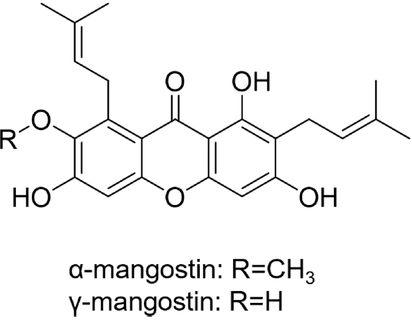
Chemical structures of α- and γ-mangostins.
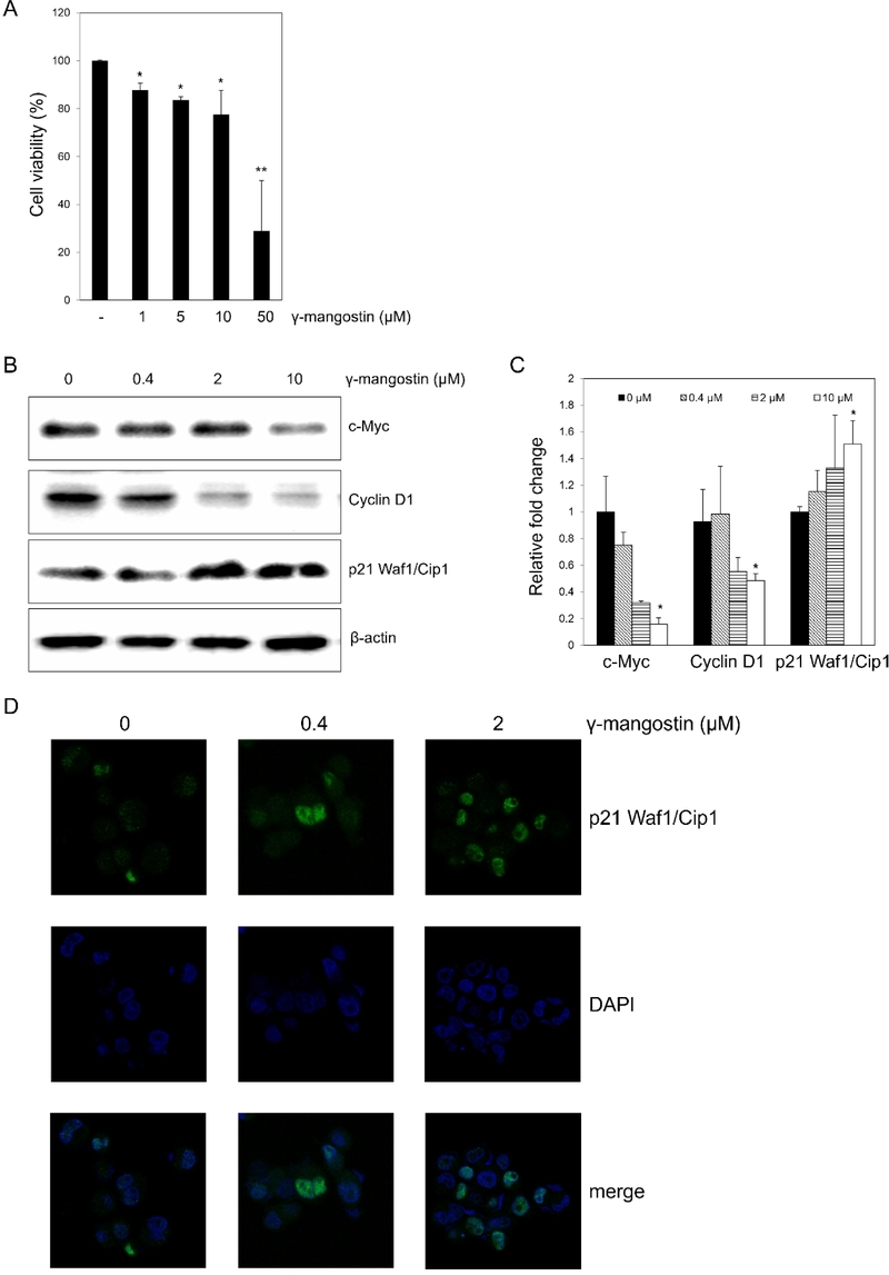
Effect of γ-mangostin on proliferation in MKN45 cells. (A) Effect of γ-mangostin on the cell viability. MKN45 cells were treated with γ-mangostin (B) The expression of c-Myc, CD44, cyclin D1 and p21 Waf1/Cip1 was assayed by western blot in cells treated with γ-mangostin (0.4, 2, and 10 µM) for 24 hours. (C) The expression of c-Myc, cyclin D1 and p21 Waf1/Cip1 was assayed by qRT-PCR in cells treated with γ-mangostin (0.4, 2, and 10 µM) for 12 hours. (D) Expression of p21 Waf1/Cip1 by γ-mangostin. The cells were cultured for 24 hours, fixed, permeabilized, and incubated with rabbit monoclonal anti-p21 Waf1/Cip1 antibody followed by Alexa 488 conjugated anti-rabbit Ig (green). The nuclei of the corresponding cells were visualized by 4′,6-diamidino-2-phenylindole (DAPI) staining.
Among the downstream targets linked to c-Met-mediated proliferation, c-Myc, cyclin D1, and p21 Waf1/Cip1 were selected for further analysis.23 γ-Mangostin produced a dose-related elevation of p21 Waf1/Cip1 together with concomitant reductions in c-Myc and cyclin D1, as presented in Fig. 2B. Similar dose-dependent modulation by γ-mangostin was observed in mRNA expression level of c-Myc, cyclin D1, and p21 Waf1/Cip1 (Fig. 2C). qRT-PCR experiments for the indicated genes in Fig. 2C showed significant reduction of c-Myc and cyclin D1 mRNA expression and significant increment of p21 Waf1/Cip1 mRNA expression with 10 μM γ-mangostin treatment. Consistent with immunoblot and qRT-PCR results in Fig. 2B and 2D, expression of p21 Waf1/Cip1 in immunofluorescence analysis increased dose-dependently with 2 μM γ-mangostin treatment (Fig. 2D).
In order to investigate mechanism of γ-mangostin on cell proliferation, effects on c-Met signaling pathways were studied. The addition of γ-mangostin for 24 h suppressed c-Met activation as shown by p-c-Met western blot with dose-dependent manner without affecting c-Met expression (Fig. 3). ERK1/2 and STAT3 activation represented by ERK and STAT3 phosphorylation level (p-ERK and p-STAT3, respectively) were also inhibited by γ-mangostin treatment. ERK1/2 and STAT3 are major downstream signaling pathways of c-Met receptor kinase. Reduction of c-Met and STAT3 phosphorylation were observed from 0.4 μM of γ-mangostin, while p-ERK blot reduction was observed from 2 μM of γ-mangostin. Taken together, these findings suggest that γ-mangostin effectively attenuates c-Met-mediated signaling cascades essential to the proliferative capacity of cancer cells
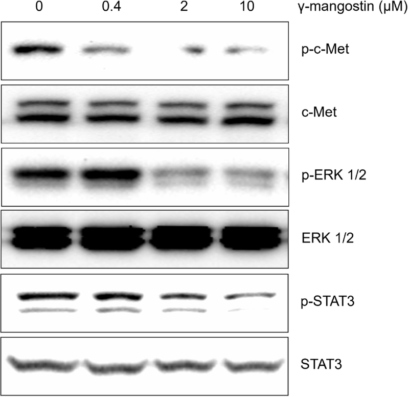
Effect of γ-mangostin on the c-Met activation and its downstream signaling pathway in the MKN45 human gastric cancer cell line. The cells were treated with γ-mangostin at various doses (0.4–10 µM) for 24 h. Western blotting experiments for c-Met, ERK1/2, STAT3 and its phosphorylated protein were performed with the cell lysates.
In order to prove that c-Met pathway mediates γ–mangostin-induced cell cytotoxicity, c-Met knockdown approach was employed. c-Met expression was knocked down by transfecting specific siRNAs against c-met gene. With c-Met siRNA transfection, c-Met phosphorylation as well as c-Met expression was decreased (Fig. 4A). 1 μM of c-Met siRNA exhibited strongest knockdown effect. Thus subsequent experiments were carried out with 1 μM concentration of c-Met siRNA. When c-Met expression was knocked down using c-Met siRNA, downregulation of c-Myc and upregulation of p21 Waf1/Cip1 by γ–mangostin were abolished (Fig. 4B and 4D). Fig. 4C shows western blot of apoptotic factors with c-Met knock-down, in the presence and absence of γ–mangostin. With c-Met siRNA treatment in the absence of γ–mangostin, increase of caspase-3 cleavage was observed. When γ–mangostin (2 μM) was treated in c-Met knock-down cells, caspase-3 cleavage was not induced and rather cleaved caspase-3 level was decreased. Collectively, the data suggest a dependence on c-Met signaling for the observed γ-mangostin-induced changes in proliferation-associated protein expression. We sought to determine whether the ability of hepatocyte growth factor (HGF) to bind HGFR to small molecules in the vicinity of binding to HGFR could prevent c-MET activation by HGF. To confirm the purpose, the protein structure16 in which HGFR is bound to HGF was obtained from PDB17 and molecular docking studies were carried out. Previous studies on mutations in HGF have affected HGFR-HGF binding when V692 of the HGF-β chain is mutated.16 With the previous studies, binding site was selected by considering D190, R191 and F192 of HGFR, which is a counterpart of HGF binding to C692, as the key residues (Fig. 5). As a result, we got the ranked pose based on CDOCKER_ENERGY, and selected the final binding mode among them. The final binding mode is shown in Fig. 6. Each compound is capable of π-anion interaction with D190 and hydrogen bond with D191 using xanthone scaffold, and formation of hydrophobic interaction via alkyl chain. When superimposing the PDB bound to HGF, which is an inherent ligand, it was confirmed that it occupies the vicinity including Y673 and V692 spatially.
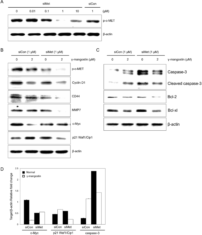
The siRNA directed against c-Met affects cell-cycle progression and apoptosis under γ-mangostin treatment. (A) MKN45 cells were transfected with siRNA c-Met at indication condition for 48 hours. Western blotting was used to determine the protein expression of p-c-Met with relative to β-actin reference protein. (B) Control siRNA and siRNA c-Met transfected MKN45 cells were treated with or without γ-mangostin (2 µM) for 24 hr. Cells were harvested to get total lysates, then performed in western blotting to observe the expression of p-c-Met, cyclin D1, CD44, MMP7, c-Myc p21 Waf1/Cip1 and β-actin (C) As same as (B) conditions, cells were harvested to get total lysates, then performed in western blotting to observe the expression of Caspase-3, Bcl-2, Bcl-xl and β-actin. (D) The immunoblot signals were quantified using Molecular Analyst/PC densitometry software (Bio-Rad). Densitometric analysis of c-Myc, p21 Waf1/Cip1 and Caspase-3 is reported. The relative quantity of the target protein of interest was then normalized to the relative quantity of β-actin.
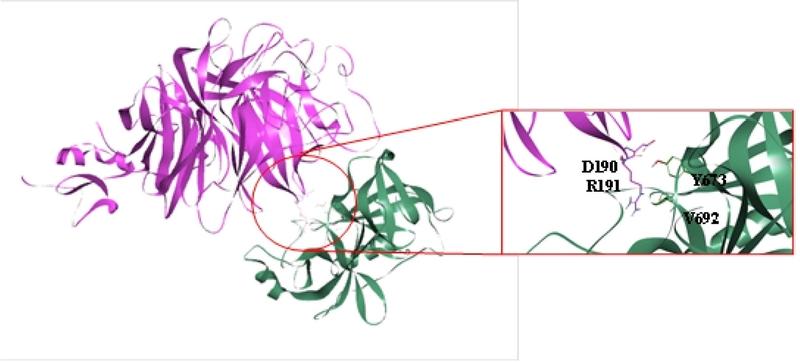
Key residues in binding site of HGFR and HGF complex. The structure is HGFR-HGF complex (PDB code: 1SHY). Pink means HGFR and green means HGF. Key residues are expressed in stick style in red box.
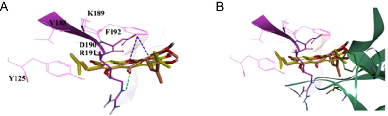
The predicted binding mode of α-mangostin and γ-mangostin. (A) The suggested binding mode of α-mangostin and γ-mangostin with HGFR. Green dashed line means hydrogen bond and blue dashed lines mean π-anion interaction. Hydrophobic interactions are not present in figure. (B) The binding mode is superimposed on the complex structure. HGFR and HGF are shown in pink and green line and ribbon style, respectively. Orange stick and yellow stick are α-mangostin and γ-mangostin, respectively.
Globally, gastric cancer represents one of the most common forms of cancer and accounts for the second-highest number of cancer-related deaths.24 Since most patients are diagnosed with metastatic disease, systemic chemotherapy serves as the primary therapeutic approach; however, prognosis remains poor, with median survival generally under one year.25 c-Met is now widely recognized as a valuable candidate for cancer-directed therapy, owing to its involvement in multiple processes driving human cancers, including tumor growth, invasive spread, and the formation of new blood vessels.12 Combining c-Met inhibitors with additional therapeutic agents could play a key role in both circumventing drug resistance and enhancing overall clinical benefit. To date, no fewer than 16 such agents have been subjected to clinical testing.26
c-Met dimer formation leads to autophosphorylation at defined tyrosine residues, which creates docking sites for the recruitment of intracellular signaling proteins responsible for initiating MAPK and STAT pathway activation.27 Selective modulation of docking proteins and downstream mediators has been used to elucidate their respective roles in regulating c-Met pathway activity.28,29 In this study, γ-mangostin was shown to reduce the levels of two downstream signaling markers – p-ERK, associated with the MAPK pathway, and p-STAT3, linked to JAK/STAT3 signaling. Consistent with this, a recent report has shown that α-mangostin inhibits gastric adenocarcinoma cell growth in vitro through the STAT3 pathway.30 Our findings likewise demonstrate that γ-mangostin attenuates c-Met-associated signaling by reducing p-ERK and p-STAT3 expression in MKN45 gastric cancer cells, suggesting that γ-mangostin exerts anticancer activity in c-Met-activated cancer cells. Notably, however, γ-mangostin did not directly inhibit c-Met kinase activity in an in vitro kinase assay (data not shown), indicating that its inhibitory effect on c-Met signaling likely occurs downstream of the receptor rather than through direct kinase inhibition.
In conclusion, γ–mangostin was found to have an inhibitory effect against MKN45 cell proliferation, but also exhibited activity to c-Myc and Cyclin D1 down-regulation. Of interest, MKN45 lacking c-Met gene, which escaped from γ–mangostin induced p21 Waf1/Cip1 expression and apoptosis. Thus, the potential anti-cancer activity of γ–mangostin may be strongly associated with the blocking of c-Met signal pathways, including anti-oncogenic pathways.
Acknowledgments
This work was supported by a grant (NRF-RS-2024-00410675) from the National Research Foundation of Korea (NRF), funded by the Korean government (MSIT).
Conflicts of Interest
The authors declare that they have no conflicts of interest.
References
-
Moongkarndi, P.; Kosem, N.; Kaslungka, S.; Luanratana, O.; Pongpan, N.; Neungton, N. J. Ethnopharmacol. 2004, 90, 161–166.
[https://doi.org/10.1016/j.jep.2003.09.048]

-
Chin, Y.-W.; Kinghorn, A. D. Mini-Rev. Org. Chem. 2008, 5, 355–364.
[https://doi.org/10.2174/157019308786242223]
-
Dharmaratne, H. R. W.; Sakagami, Y.; Piyasena, K. G. P.; Thevanesam, V. Nat. Prod. Res. 2013, 27, 938–941.
[https://doi.org/10.1080/14786419.2012.678348]
-
Yoo, J.-H.; Kang, K.; Jho, E. H.; Chin, Y.-W.; Kim, J.; Nho, C. W. Food Chem. 2011, 129, 1559–1566.
[https://doi.org/10.1016/j.foodchem.2011.06.007]
-
Chen, L.-G.; Yang, L.-L.; Wang, C.-C. Food Chem. Toxicol. 2008, 46, 688–693.
[https://doi.org/10.1016/j.fct.2007.09.096]
-
Widowati, W.; Darsono, L.; Suherman, J.; Fauziah, N.; Maesaroh, M.; Erawijantari, P. P. Nat. Prod. Sci. 2016, 22, 147–153.
[https://doi.org/10.20307/nps.2016.22.3.147]
-
Chae, H.-S.; Oh, S.-R.; Lee, H.-K.; Joo, S. H.; Chin, Y.-W. Food Chem. 2012, 134, 397–400.
[https://doi.org/10.1016/j.foodchem.2012.02.075]
-
Peruzzi, B.; Bottaro, D. P. Clin. Cancer Res. 2006, 12, 3657–3660.
[https://doi.org/10.1158/1078-0432.CCR-06-0818]
-
Cooper, C. S.; Park, M.; Blair, D. G.; Tainsky, M. A.; Huebner, K.; Croce, C. M.; Vande Woude, G. F. Nature 1984, 311, 29–33.
[https://doi.org/10.1038/311029a0]
-
Birchmeier, C.; Birchmeier, W.; Gherardi, E.; Vande Woude, G. F. Nat. Rev. Mol. Cell Biol. 2003, 4, 915–925.
[https://doi.org/10.1038/nrm1261]
-
Comoglio, P. M.; Giordano, S.; Trusolino, L. Nat. Rev. Drug Discov. 2008, 7, 504–516.
[https://doi.org/10.1038/nrd2530]
-
Christensen, J. G.; Burrows, J.; Salgia, R. Cancer Lett. 2005, 225, 1–26.
[https://doi.org/10.1016/j.canlet.2004.09.044]
-
Engelman, J. A.; Zejnullahu, K.; Mitsudomi, T.; Song, Y.; Hyland, C.; Park, J. O.; Lindeman, N.; Gale, C.-M.; Zhao, X.; Christensen, J.; Kosaka, T.; Holmes, A. J.; Rogers, A. M.; Cappuzzo, F.; Mok, T.; Lee, C.; Johnson, B. E.; Cantley, L. C.; Jänne, P. A. Science 2007, 316, 1039–1043.
[https://doi.org/10.1126/science.1141478]
-
Shan, T.; Ma, Q.; Guo, K.; Liu, J.; Li, W.; Wang, F.; Wu, E. Curr. Mol. Med. 2011, 11, 666–677.
[https://doi.org/10.2174/156652411797536679]
-
Matsumoto, K.; Akao, Y.; Ohguchi, K.; Ito, T.; Tanaka, T.; Iinuma, M.; Nozawa, Y. Bioorg. Med. Chem. 2005, 13, 6064–6069.
[https://doi.org/10.1016/j.bmc.2005.06.065]
-
Stamos, J.; Lazarus, R. A.; Yao, X.; Kirchhofer, D.; Wiesmann, C. EMBO J. 2004, 23, 2325–2335.
[https://doi.org/10.1038/sj.emboj.7600243]
-
Berman, H. M.; Battistuz, T.; Bhat, T. N.; Bluhm, W. F.; Bourne, P. E.; Burkhardt, K.; Feng, Z.; Gilliland, G. L.; Iype, L.; Jain, S.; Fagan, P.; Marvin, J.; Padilla, D.; Ravichandran, V.; Schneider, B.; Thanki, N.; Weissig, H.; Westbrook, J.; Zardecki, C. Acta Crystallogr. D Biol. Crystallogr. 2002, 58, 899–907.
[https://doi.org/10.1107/S0907444902003451]
- Creighton, T. E. Proteins: Structures and Molecular Properties; W.H. Freeman: New York, 1993.
- Dassault Systèmes. BIOVIA Discovery Studio Simulation. https://discover.3ds.com/ko/biovia-discovery-studio-simulation, (accessed July 3, 2026).
-
Groom, C. R.; Bruno, I. J.; Lightfoot, M. P.; Ward, S. C. Acta Crystallogr. B Struct. Sci. Cryst. 2016, 72, 171–179.
[https://doi.org/10.1107/S2052520616003954]
-
Wu, G.; Robertson, D. H.; Brooks III, C. L.; Vieth, M. J. Comput. Chem. 2003, 24, 1549–1562.
[https://doi.org/10.1002/jcc.10306]
-
Momany, F. A.; Rone, R. J. Comput. Chem. 1992, 13, 888–900.
[https://doi.org/10.1002/jcc.540130714]
-
Danilkovitch-Miagkova, A.; Zbar, B. J. Clin. Invest. 2002, 109, 863–867.
[https://doi.org/10.1172/JCI0215418]
-
Ferlay, J.; Shin, H.-R.; Bray, F.; Forman, D.; Mathers, C.; Parkin, D. M. Int. J. Cancer 2010, 127, 2893–2917.
[https://doi.org/10.1002/ijc.25516]
-
Pyrhönen, S.; Kuitunen, T.; Nyandoto, P.; Kouri, M. Br. J. Cancer 1995, 71, 587–591.
[https://doi.org/10.1038/bjc.1995.114]
-
Liu, X.; Newton, R. C.; Scherle, P. A. Trends Mol. Med. 2010, 16, 37–45.
[https://doi.org/10.1016/j.molmed.2009.11.005]
-
Comoglio, P. M. Nat. Cell Biol. 2001, 3, E161–E162.
[https://doi.org/10.1038/35083116]
-
Furge, K. A.; Zhang, Y.-W.; Vande Woude, G. F. Oncogene 2000, 19, 5582–5589.
[https://doi.org/10.1038/sj.onc.1203859]
-
Wong, A. S. T.; Roskelley, C. D.; Pelech, S.; Miller, D.; Leung, P. C. K.; Auersperg, N. Exp. Cell Res. 2004, 299, 248–256.
[https://doi.org/10.1016/j.yexcr.2004.06.002]
-
Shan, T.; Cui, X.; Li, W.; Lin, W.; Lu, H.; Li, Y.; Chen, X.; Wu, T. Acta Pharmacol. Sin. 2014, 35, 1065–1073.
[https://doi.org/10.1038/aps.2014.43]