
Phytochemical Constituents of the Aerial Parts of Artemisia iwayomogi Kitamura and Their Anti-inflammatory Activities
Abstract
Artemisia iwayomogi Kitamura is a well-known medicinal plant widely utilized in traditional medicine for its various pharmacological properties. In this study, phytochemical investigation of the aerial parts of A. iwayomogi led to the isolation and characterization of 13 secondary metabolites, including one new (1) and 12 known compounds (2–13). The structural elucidation of the new compound was achieved through comprehensive spectroscopic analysis, including NMR and HRMS data, and a modified Mosher’s method. This study represents the first report of compounds 2, 3, 5, and 13 from this plant species. Compound 1 was evaluated for its inhibitory activity against NO production in LPS-stimulated macrophages. However, it was found to be inactive against NO production (IC50 > 100 μM). In silico target prediction using SwissTargetPrediction identified catechol O-methyltransferase (COMT) as the highest-probability predicted target of compound 1, providing a hypothesis-generating basis for future biological investigation.
Keywords:
Artemisia iwayomogi, Anti-inflammatory, NMR, In silicoIntroduction
The nitric oxide synthase (NOS) family of enzymes comprises the constitutive isoforms endothelial NOS (eNOS) and neuronal NOS (nNOS), along with the inducible form (iNOS). These enzymes are responsible for the synthesis of nitric oxide (NO), an essential intercellular signaling molecule, from L-arginine.1,2 Constitutive nitric oxide production under physiological conditions plays a crucial role in supporting innate immune defense against pathogens, modulating neuronal communication, and maintaining vascular homeostasis through the process of vasodilation.2 NO exhibits concentration-dependent biphasic effects. While basal levels of NO are cytoprotective, excessive production, particularly through iNOS induction in response to inflammatory triggers such as lipopolysaccharide (LPS) and pro-inflammatory cytokines, can initiate pathological cascades. Excess NO rapidly reacts with superoxide anions to form peroxynitrite (ONOO⁻), a potent reactive nitrogen species that induces DNA damage, lipid peroxidation, protein nitration, and cellular dysfunction in oxidative conditions.2,3 Numerous diseases, including cancer, inflammatory bowel disease, cardiovascular conditions, septic shock, and neurodegenerative disorders, are affected by oxidative and nitrosative stress.2 Selective inhibitors of iNOS have emerged as promising therapeutic candidates because of the enzyme’s involvement in various pathologies. Natural products serve as an excellent resource for discovering novel NO-modulating agents due to their structural diversity and ability to interact with multiple targets.
Known locally as “Dowijigi” or “Haninjin” in Korea, Artemisia iwayomogi Kitamura is a perennial herb that is part of the Asteraceae family. This plant has a long history of use in traditional East Asian medicine, where it has been employed to address a range of ailments, particularly those affecting the liver, such as cirrhosis and hepatitis, along with various inflammatory and immunomodulatory disorders.4,5 Numerous bioactive secondary metabolites present in the plant have been identified through comprehensive phytochemical analysis. These encompass coumarins such as scopoletin and scopolin, flavonoids including genkwanin and jaceosidin, as well as phenolic acids like chlorogenic acid.4,6 Pharmacological investigations have demonstrated significant anti-inflammatory effects by inhibiting pro-inflammatory cytokines (TNF-α, IL-6) through the downregulation of p38 MAPK and NF-κB signaling pathways, which are crucial transcriptional regulators of iNOS production.4 Moreover, compounds like chlorogenic acid and scopoletin demonstrate direct scavenging ability against ONOO⁻, in addition to possessing anti-allergic, bone metabolism-regulating, and antimicrobial characteristics.6,7 Recently, we reported the isolation and anti-inflammatory activity of several compounds from A. iwayomogi.5 Given the plant’s rich phytochemical diversity and established anti-inflammatory properties, continued investigation of its secondary metabolites for NO inhibitory potential is warranted. Herein, we describe the isolation and structural elucidation of additional compounds from A. iwayomogi and their inhibitory effects on NO production in LPS-induced RAW 264.7 macrophages.
Experimental
General experimental procedures – Optical rotations were measured using a JASCO P-2000 digital polarimeter. NMR spectra were acquired using the Varian Unity Inova 400 MHz spectrometer (Varian, Inc., California, USA). High-resolution electron impact-mass spectrometry (HREIMS) was carried out using a JEOL JMS-AX 300L spectrometer (Jeol, Akishima, Tokyo, Japan). Thin-layer chromatography (TLC) was conducted utilizing RP-18 F254s and Merck precoated silica gel F254 plates. Column chromatography (CC) utilized (RP)-C18 (Merck, 75 mesh) and silica gel 60 (Merck, 230–400 mesh). Preparative HPLC was performed utilizing a YMC Pak ODS-A column (20 × 250 mm, 5 μm particle size; YMC Co., Ltd., Japan) and a Waters 2487 controller system equipped with a UV detector (UV/VIS-156).
Plant material – The aerial parts of A. iwayomogi were collected at the Medicinal Plants Garden at Daegu Catholic University in October 2020 and authenticated by Professor Byung Sun Min, one of the authors. A voucher specimen (voucher no. CUD-3030) was deposited at the Laboratory of Pharmacognosy in the College of Pharmacy, Daegu Catholic University, Republic of Korea.
Extraction and isolation – The chopped, air-dried aerial parts of A. iwayomogi (7.3 kg) underwent exhaustive extraction with 100% MeOH (3 × 10 L). The resulting filtrates were pooled and evaporated under reduced pressure using a vacuum rotary evaporator, yielding 750 g of a crude extract. This residue was subsequently suspended in distilled water and sequentially partitioned with n-hexane, dichloromethane (MC), ethyl acetate (EA), and n-butanol (n-BuOH), respectively. The EA extract (150 g) was fractionated via silica gel column chromatography (CC), employing a gradient system of CH2Cl2:MeOH (1:0–0:1, v/v) to afford 15 subfractions (Fr.1–Fr.15). Fr.3 (1.4 g) was chromatographed on a silica gel column using a n-hexane:acetone gradient (3:1–0:1) to provide nine subfractions (Fr.3.1–Fr.3.9). Fr.3.2 (118.8 mg) was purified by preparative HPLC (MeOH:H2O, 20:80–80:20, 5 mL/min) to yield compounds 2 (1.0 mg), 3 (1.3 mg), 10 (11.5 mg), and 11 (3.3 mg). Fr.3.5 (455.1 mg) was separated by Sephadex LH-20 to obtain 9 (2.1 mg), 12 (7.3 mg), and 13 (5.6 mg). Fr.3.6 (90.0 mg) was purified by preparative HPLC (MeOH:H2O, 40:60–70:30, 5 mL/min) to yield compounds 1 (5.5 mg), 4 (3.8 mg), and 8 (2.5 mg). Further elution of Fr.3.8 (164.3 mg) through an RP-C18 column (MeOH:H2O, 1:2–3:1) afforded compounds 5 (3.6 mg), 6 (8.3 mg), and 7 (4.1 mg).
Artemisiwayin (1) – White amorphous powder; [α] +17.5 (c 0.015, MeOH); 1H (400 MHz, methanol-d4) and 13C NMR (100 MHz, methanol-d4) data, see Table 1; HREIMS m/z 182.0577 [M]+ (calcd for C9H10O4, 182.0579).

1H (400 MHz) and 13C (100 MHz) NMR data in methanol-d4 for compound 1.
p-Coumaric acid (2) – 1H NMR (400 MHz, methanol-d4) δH 7.61 (1H, d, J = 15.8 Hz), 7.46 (2H, d, J = 8.7 Hz), 6.81 (2H, d, J = 8.7 Hz), 6.29 (1H, d, J = 15.8 Hz); 13C NMR (100 MHz, methanol-d4) δC 169.6, 159.7, 145.2, 129.6, 125.8, 115.4, 114.2.
Caffeic acid (3) – 1H NMR (400 MHz, methanol-d4) δH 7.48 (1H, d, J = 15.8 Hz), 6.98 (1H, m), 6.88 (1H, m), 6.72 (1H, m), 6.16 (1H, d, J = 15.8 Hz); 13C NMR (100 MHz, methanol-d4) δC 171.1, 149.4, 147.0, 146.7, 127.8, 122.8, 116.5, 115.5, 115.1.
Methyl caffeate (4) – 1H NMR (400 MHz, methanol-d4) δH 7.56 (1H, d, J = 15.8 Hz), 7.05 (1H, s), 6.95 (1H, d, J = 7.8 Hz), 6.79 (1H, d, J = 7.8 Hz), 6.27 (1H, d, J = 15.8 Hz), 3.77 (3H, s); 13C NMR (100 MHz, methanol-d4) δC 169.7, 149.5, 146.9, 146.8, 127.7, 122.9, 116.5, 115.1, 114.8, 52.0.
Hydroquinone (5) – 1H NMR (400 MHz, methanol-d4) δH 6.62 (4H, s); 13C NMR (100 MHz, methanol-d4) δC 151.2, 116.8.
4-Hydroxybenzoic acid (6) – 1H NMR (400 MHz, methanol-d4) δH 7.90 (2H, d, J = 8.8 Hz), 6.84 (2H, d, J = 8.8 Hz); 13C NMR (100 MHz, methanol-d4) δC 170.0, 163.3, 133.0, 122.7, 116.0.
Protocatechuic acid (7) – 1H NMR (400 MHz, methanol-d4) δH 7.45 (1H, s), 7.40 (1H, d, J = 8.2 Hz), 6.78 (1H, d, J = 8.2 Hz); 13C NMR (100 MHz, methanol-d4) δC 173.4, 150.1, 145.6, 127.2, 123.4, 117.8, 115.5.
Eugenyl glucoside (8) – 1H NMR (400 MHz, methanol-d4) δH 7.08 (1H, d, J = 8.2 Hz), 6.82 (1H, d, J = 1.9 Hz), 6.72 (1H, dd, J = 8.2, 1.9 Hz), 5.95 (1H, ddt, J = 16.8, 10.0, 6.7 Hz), 5.02 (1H, m), 4.85 (1H, d, J = 7.4 Hz), 3.87 (1H, dd, J = 12.1, 1.3 Hz), 3.83 (3H, s), 3.69 (1H, dd, J = 12.1, 5.1 Hz), 3.35–3.49 (overlap); 13C NMR (100 MHz, methanol-d4) δC 150.7, 146.3, 139.0, 136.4, 122.1, 118.3, 115.8, 114.1, 103.0, 78.1, 77.8, 74.9, 71.3, 62.5, 56.7, 40.7.
Kaempferol (9) – 1H NMR (400 MHz, DMSO-d6) δH 8.04 (2H, d, J = 8.8 Hz), 6.93 (2H, d, J = 8.8 Hz), 6.44 (1H, d, J = 1.97 Hz), 6.20 (1H, d, J = 1.97 Hz); 13C NMR (100 MHz, DMSO-d6) δC 175.8, 163.8, 160.7, 160.5, 159.1, 156.2, 146.7, 135.6, 129.5, 121.7, 115.4, 103.0, 98.1, 93.5.
Luteolin (10) – 1H NMR (400 MHz, methanol-d4) δH 7.34 (1H, overlap), 7.32 (1H, overlap), 6.88 (1H, d, J = 7.2 Hz), 6.49 (1H, s), 6.40 (1H, s), 6.18 (1H, s); 13C NMR (100 MHz, methanol-d4) δC 183.8, 166.3, 165.9, 163.1, 159.3, 150.9, 146.9, 123.6, 120.3, 116.8, 114.2, 105.3, 103.8, 100.1, 95.0.
Apigenin (11) – 1H NMR (400 MHz, DMSO-d6) δH 7.91 (2H, d, J = 8.7 Hz), 6.92 (2H, d, J = 8.7 Hz), 6.76 (1H, s), 6.47 (1H, brs), 6.19 (1H, brs); 13C NMR (100 MHz, DMSO-d6) δC 181.7, 164.1, 163.7, 161.4, 161.2, 157.3, 128.4, 121.2, 115.9, 103.7, 102.8, 98.8, 94.0.
Naringenin (12) – 1H NMR (400 MHz, methanol-d4) δH 7.33 (2H, d, J = 8.4 Hz), 6.84 (2H, d, J = 8.4 Hz), 5.91 (2H, overlap), 5.35 (1H, dd, J = 12.9, 2.8 Hz), 3.12 (1H, dd, J = 17.1, 13.0 Hz), 2.71 (1H, dd, J = 17.1, 3.0 Hz); 13C NMR (100 MHz, methanol-d4) δC 197.8, 168.3, 165.5, 164.9, 159.0, 131.1, 129.0, 116.3, 103.4, 97.0, 96.2, 80.5, 44.0.
Eriodictyol (13) – 1H NMR (400 MHz, acetone-d6) δH 7.04 (1H, s), 6.88 (2H, s), 5.97 (1H, brs), 5.96 (1H, brs), 5.40 (1H, dd, J = 12.7, 3.0 Hz), 3.14 (1H, dd, J = 17.1, 12.7 Hz), 2.73 (1H, dd, J = 17.1, 3.0 Hz); 13C NMR (100 MHz, acetone-d6) δC 197.2, 167.3, 165.3, 165.0, 164.3, 146.3, 146.0, 131.6, 119.3, 116.0, 114.7, 103.2, 96.8, 95.8, 79.9, 43.6.
Determination of absolute configuration – Compound 1 (1.0 mg) was treated separately with (R)- and (S)-MTPA-Cl in pyridine. The mixtures were stirred at room temperature until the reactions were complete to yield the (S)- and (R)-MTPA esters, respectively. After that, 1H NMR spectra were recorded, and the absolute configuration was assigned based on the sign distribution of ΔδH(S-R) values following the modified Mosher’s method.8
Determination of NO production and cell viability – RAW 264.7 cells were exposed to either 1 μg/mL of LPS (Sigma Chemical Co.) or a control for 24 hours, with or without the test substance (1–100 μM). A total of 100 μL of Griess reagent (consisting of 0.1% naphthyl ethylenediamine dihydrochloride in distilled water and 1% sulfanilamide in 5% phosphoric acid) was combined with 100 μL of the cell culture supernatant following incubation. Cell viability was evaluated via an MTT-based colorimetric test.9
In silico target prediction – Biological targets for compound 1 were predicted using the SwissTargetPrediction webserver (http://www.swisstargetprediction.ch/) against Homo sapiens. The structure was submitted as a SMILES string to identify potential interactions with protein classes based on 2D/3D structural similarity to known ligands.10
Results and Discussion
The methanolic extract from the aerial parts of A. iwayomogi was sequentially partitioned using n-hexane, MC, EA, and n-BuOH. Intensive fractionation of the EA-soluble residue (150 g) via silica gel chromatography and preparative HPLC yielded one new (1), and 12 known compounds (2–13) (Fig. 1), which were determined to be p-coumaric acid (2),11 caffeic acid (3),12 methyl caffeate (4),12 hydroquinone (5),13 4-hydroxybenzoic acid (6),14 protocatechuic acid (7),15 eugenyl glucoside (8),16 kaempferol (9),17 luteolin (10),18 apigenin (11),19 naringenin (12),20 and eriodictyol (13)21 by analyzing and comparing their spectroscopic data with those reported in the literature. Notably, this study marks the first isolation of compounds 2, 3, 5, and 13 from this plant species.
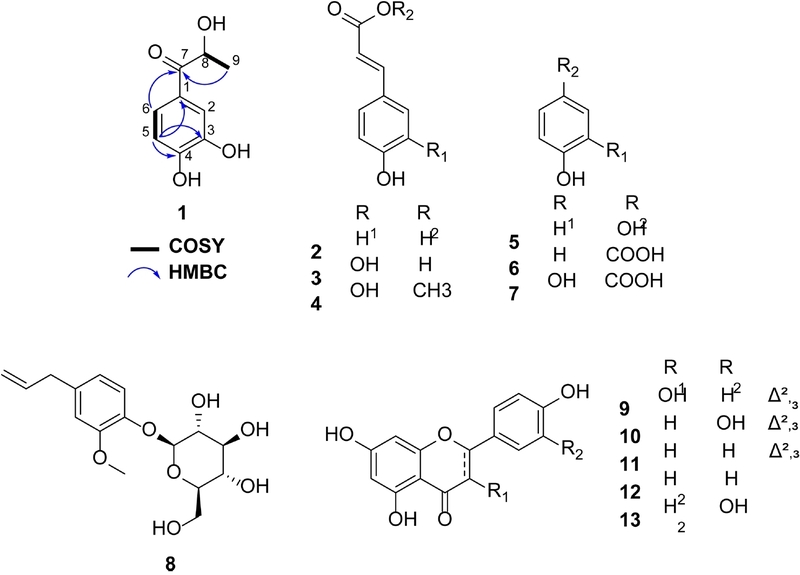
Chemical structures of compounds 1–13 isolated from A. iwayomogi and key COSY and HMBC correlations for the new compound 1
Artemisiwayin (1) was isolated as a white amorphous powder with a molecular formula of C9H10O4, which was deduced from the HREIMS that displayed a molecular ion peak at m/z 182.0577 [M]+ (calcd for C9H10O4, 182.0579). Its 1H NMR spectrum (Table 1) revealed an ABX coupling system characterized by signals at δH 7.43 (2H, overlap, H-2 and H-6) and 6.84 (1H, d, J = 8.8 Hz, H-5), along with one methine signal at δH 5.11 (1H, q, J = 6.9 Hz, H-8) and one methyl signal at δH 1.39 (3H, d, J = 6.9 Hz, H-9). The 13C NMR spectrum displayed 9 carbon signals, including one carbonyl (δC 202.1), six aromatic carbons (δC 152.5, 146.6, 127.6, 123.5, 116.4, 116.0), one oxygenated methine (δC 69.9), and one methyl (δC 22.2). Comparison with 1-(3,4-dihydroxyphenyl)-1-hydroxy-2-propanone revealed close structural similarities, with the key difference being the positions of the carbonyl and hydroxy groups.22 In compound 1, these groups were located at C-7 and C-8, respectively, whereas in the reference compound, they were at C-8 and C-7. The structural assignment was confirmed by HMBC correlations from H-6 to C-7 and H-9 to both C-8 and C-7, as well as a COSY cross-peak between H-9 and H-8 (Fig. 1). Based on these findings, a planar structure was proposed for compound 1. The absolute configuration at C-8 of 1 was determined through application of the modified Mosher’s method.8 Treatment of compound 1 with (R)- and (S)-MTPA-Cl independently yielded the corresponding (S)-and (R)-MTPA esters, respectively. Analysis of the ΔδH(S-R) values revealed a positive value for H-5 and a negative value for H3-9, thereby assigning the S configuration to C-8 (Fig. 2). Therefore, the structure of 1 was deduced and given the trivial name artemisiwayin.
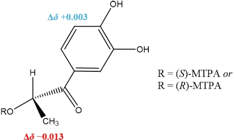
ΔδH(S-R) values obtained from the 1H NMR spectra of the MTPA esters of compound 1.
Compound 2 was identified as p-coumaric acid. The 1H NMR spectrum exhibited characteristic signals for a para-disubstituted benzene ring at δH 7.46 (2H, d, J = 8.7 Hz, H-2/6) and 6.81 (2H, d, J = 8.7 Hz, H-3/5). The presence of a trans-configured olefinic side chain was confirmed by two coupled doublets at δH 7.61 (1H, d, J = 15.8 Hz, H-7) and 6.29 (1H, d, J = 15.8 Hz, H-8). The 13C NMR spectrum displayed seven signals, including a carbonyl carbon at δC 169.6 (C-9), an oxygenated aromatic carbon at δC 159.7 (C-4), and two olefinic carbons at δC 145.2 (C-7) and 114.2 (C-8). These spectroscopic data were in full agreement with literature values for p-coumaric acid.11
The NMR spectra of 3 were closely related to those of 2, except for the aromatic region, which exhibited a 1,2,4-trisubstituted benzene system (δH 6.98, 6.88, and 6.72) instead of the para-disubstituted pattern found in 2. The presence of two oxygenated aromatic carbons at δC 147.0 (C-3) and 146.7 (C-4) further confirmed the catechol moiety. These spectroscopic data were consistent with literature values for caffeic acid.12
Compound 5 was identified as hydroquinone. The NMR spectra revealed a high degree of structural symmetry. The 1H NMR spectrum exhibited a single resonance at δH 4.85 (4H, s), corresponding to the four equivalent aromatic protons. Consistently, the 13C NMR spectrum displayed only two signals: one oxygenated aromatic carbon at δC 151.2 (C-1/4) and one aromatic carbon at δC 116.8 (C-2/3/5/6). These spectroscopic characteristics were in complete agreement with the structure of hydroquinone.13
The NMR data for 13 were diagnostic of a flavanone skeleton. The 1H NMR spectrum displayed a typical ABX system for the C-ring at δH 7.04 (1H, s) and 6.88 (2H, s). The A-ring substitution pattern was evidenced by two meta-coupled aromatic signals at δH 5.97 and 5.96 (each 1H, brs, H-6 and H-8). In the 13C NMR spectrum, the presence of a flavanone skeleton was further confirmed by the carbonyl resonance at δC 197.2 (C-4), along with aliphatic signals at δC 79.9 (C-2) and 43.6 (C-3). Comparison of these data with literature values confirmed the identity of 13 as eriodictyol.21
To assess the anti-inflammatory potential of the isolated constituents, their ability to inhibit NO production was considered. Given that compounds 2–13 are well-known metabolites with established inhibitory profiles in the literature, further testing was focused exclusively on the new compound 1. However, compound 1 was found to be inactive under the tested conditions, with an IC50 value exceeding 100 μM. Cell viability was assessed in parallel with the NO inhibition assay across the same concentration range, and compound 1 showed no cytotoxic effect (IC50 > 100 μM).
To further elucidate the pharmacological potential of the new compound 1, an in silico target prediction analysis was performed using the SwissTargetPrediction platform. SwissTargetPrediction returned 15 putative targets, of which six had a non-zero probability score, spanning four target classes (kinase, enzyme, transferase, phosphatase) (Fig. 3, Table 2). Catechol O-methyltransferase (COMT) received the highest probability score; a further five targets, including insulin-like growth factor I receptor (IGF1R), shared an identical, lower score. The remaining nine targets (Table 2, entries 7–15) were assigned a probability of zero and are reported for completeness only, without implying a supported interaction. No NOS-family target was identified among the predictions, which may be in line with the lack of NO-inhibitory activity observed for compound 1. These results are hypothesis-generating; experimental validation, particularly against COMT, would be needed before any predicted interaction can be considered biologically relevant.
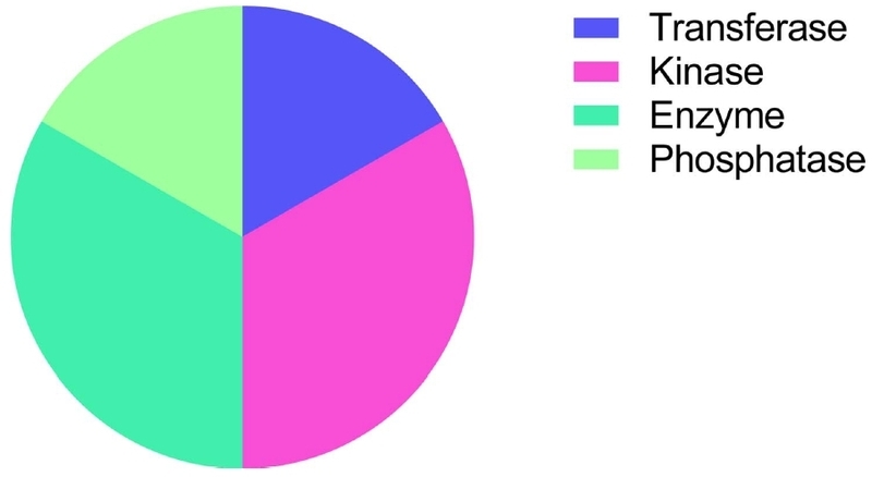
In silico target prediction of compound 1 using SwissTargetPrediction.
List of the top 15 predicted protein targets ranked by probability.*
In conclusion, 13 compounds were successfully isolated from the aerial parts of A. iwayomogi through various chromatographic techniques. The structural elucidation was confirmed by detailed spectroscopic analysis, identifying one new metabolite along with 12 known compounds. Notably, four of these (2, 3, 5, and 13) were reported for the first time from this plant source. Although compound 1 was found to be inactive against NO production (IC50 > 100 μM), the comprehensive characterization of these isolates enhances our knowledge of the plant’s chemical constituents. This study provides a scientific basis for the further exploration of A. iwayomogi as a source of diverse natural products, and future research should focus on evaluating the potential of compound 1 in other biological models or alternative therapeutic targets.
Acknowledgments
This research was supported by the ANCHOR program through the Gyeongbuk ANCHOR CENTER, funded by the Ministry of Education (MOE) and the Gyeongsangbuk-do, Republic of Korea (2026-ANCHOR-15-107).
Conflicts of Interest
The authors declare no competing financial interest.
References
-
Joo, T.; Sowndhararajan, K.; Hong, S.; Lee, J.; Park, S.-Y.; Kim, S.; Jhoo, J.-W. Saudi J. Biol. Sci. 2014, 21, 427–435.
[https://doi.org/10.1016/j.sjbs.2014.04.003]

-
Serreli, G.; Deiana, M. Antioxidants 2023, 12, 147.
[https://doi.org/10.3390/antiox12010147]
-
Jeelani, M.; Fouotsa, H.; Mohammed, O. A.; Alfaifi, J.; Adebayo, S.; Ahmed, M. M.; Yahia, A. I. O.; Eissa, H.; Bahashwan, E.; Mohammed, N. A. Front. Pharmacol. 2024, 15, 1370073.
[https://doi.org/10.3389/fphar.2024.1370073]
-
Kim, S.-H.; Choi, C.-H.; Kim, S.-Y.; Eun, J.-S.; Shin, T.-Y. Exp. Biol. Med. 2005, 230, 82–88.
[https://doi.org/10.1177/153537020523000111]
-
Vu, N. K.; Le, T. T.; Tran, T. T.; Ha, M. T.; Kim, J. A.; Min, B. S. Nat. Prod. Sci. 2025, 31, 43–48.
[https://doi.org/10.20307/nps.2025.31.1.43]
-
Kim, A. R.; Zou, Y. N.; Park, T. H.; Shim, K. H.; Kim, M. S.; Kim, N. D.; Kim, J. D.; Bae, S. J.; Choi, J. S.; Chung, H. Y. Phytother. Res. 2004, 18, 1–7.
[https://doi.org/10.1002/ptr.1358]
-
Lee, S.-H.; Ding, Y.; Yan, X. T.; Kim, Y.-H.; Jang, H.-D. J. Nat. Prod. 2013, 76, 615–620.
[https://doi.org/10.1021/np300824h]
-
Seco, J. M.; Quinoá, E.; Riguera, R. Chem. Rev. 2004, 104, 17–118.
[https://doi.org/10.1021/cr000665j]
-
Vu, N. K.; Le, T. T.; Woo, M. H.; Min, B. S. Nat. Prod. Sci. 2021, 27, 176–182.
[https://doi.org/10.20307/nps.2021.27.3.176]
-
Daina, A.; Michielin, O.; Zoete, V. Nucleic Acids Res. 2019, 47, W357–W364.
[https://doi.org/10.1093/nar/gkz382]
-
Li, Z.-J.; Cai, L.; Mei, R.-F.; Dong, J.-W.; Li, S.-Q.; Yang, X.-Q.; Zhou, H.; Yin, T.-P.; Ding, Z.-T. Tetrahedron Lett. 2015, 56, 7197–7200.
[https://doi.org/10.1016/j.tetlet.2015.11.050]
-
Umereweneza, D.; Atilaw, Y.; Peintner, S.; Rudenko, A.; Bourgard, C.; Xiong, R.; Muhizi, T.; Sunnerhagen, P.; Gogoll, A.; Erdelyi, M. Eur. J. Org. Chem. 2023, 26, e202201280.
[https://doi.org/10.1002/ejoc.202201280]
-
Ni, J.-C.; Shi, J.-T.; Tan, Q.-W.; Chen, Q.-J. Molecules 2017, 22, 2107.
[https://doi.org/10.3390/molecules22122107]
-
Ngo, Q.-M. T.; Ha, M. T.; Vu, N. K.; Kim, J. A.; Woo, M. H.; Min, B. S. Biochem. Syst. Ecol. 2021, 94, 104221.
[https://doi.org/10.1016/j.bse.2020.104221]
-
Yang, Z.-Y.; Kuboyama, T.; Kazuma, K.; Konno, K.; Tohda, C. J. Nat. Prod. 2015, 78, 2297–2300.
[https://doi.org/10.1021/acs.jnatprod.5b00290]
-
Lone, B. A.; Bhushan, A.; Ganjoo, A.; Katoch, M.; Gairola, S.; Gupta, P.; Babu, V. Nat. Prod. Res. 2023, 37, 535–541.
[https://doi.org/10.1080/14786419.2022.2066101]
-
Vu, N. K.; Kim, C. S.; Ha, M. T.; Ngo, Q.-M. T.; Park, S. E.; Kwon, H.; Lee, D.; Choi, J. S.; Kim, J. A.; Min, B. S. J. Agric. Food Chem. 2020, 68, 8797–8811.
[https://doi.org/10.1021/acs.jafc.0c02122]
-
Leiss, K. A.; Cristofori, G.; van Steenis, R.; Verpoorte, R.; Klinkhamer, P. G. Phytochemistry 2013, 93, 63–70.
[https://doi.org/10.1016/j.phytochem.2013.03.011]
-
Teles, Y. C.; Horta, C. C. R.; Agra, M. d. F.; Siheri, W.; Boyd, M.; Igoli, J. O.; Gray, A. I.; Souza, M. d. F. V. d. Molecules 2015, 20, 20161–20172.
[https://doi.org/10.3390/molecules201119685]
-
Kulić, Ž.; Steiner, V. J.; Butterer, A. Planta Med. 2026, 92, 58–80.
[https://doi.org/10.1055/a-2706-7473]
-
Buranasudja, V.; Muangnoi, C.; Sanookpan, K.; Halim, H.; Sritularak, B.; Rojsitthisak, P. Nutrients 2022, 14, 2553.
[https://doi.org/10.3390/nu14122553]
-
Wu, P.; Luo, D.; Wang, Y.; Shang, X.; Wang, B.; Deng, X.; Yuan, J. Biodes. Res. 2024, 6, 0048.
[https://doi.org/10.34133/bdr.0048]