
α- and γ-Mangostins Suppress KRAS-Associated Signaling Pathways and Inhibit the Growth of Human Lung Cancer Cells
Abstract
Constitutive activation of multiple downstream survival pathways is a hallmark of KRAS-mutant non-small cell lung cancer (NSCLC), which often leads to poor responsiveness to current therapeutic strategies. In this study, we investigated the anticancer effects of α-mangostin and γ-mangostin, xanthone compounds isolated from Garcinia mangostana L., in A549 human lung cancer cells. Both compounds inhibited A549 cell proliferation, with γ-mangostin showing greater potency than α-mangostin. Notably, α- and γ-mangostin retained comparable anti-proliferative activity in paclitaxel- and gemcitabine-resistant A549 cells, suggesting their potential effectiveness against chemo-resistant lung cancer cells. Mechanistically, both compounds suppressed multiple KRAS-associated downstream signaling pathways, including PI3K/AKT/mTOR, ERK, and STAT3 signaling. α- and γ-Mangostin downregulated phosphorylation of Akt, mTOR, 4E-BP1, p70S6K, ERK, and STAT3, but upregulated AMPK phosphorylation. These signaling changes were accompanied by suppressed expression of the proliferation-related proteins cyclin D1 and PCNA. Furthermore, α- and γ-mangostin attenuated EGF-induced Akt activation, indicating potential relevance to growth factor-driven signaling processes involved in resistance to targeted therapies. Collectively, these findings indicate that α-mangostin and γ-mangostin exhibit anti-proliferative effects through multi-target inhibition of oncogenic signaling networks and may have therapeutic potential for KRAS-mutant and drug-resistant NSCLC.
Keywords:
Mangostin, Lung cancer, Anti-proliferation, Chemo-resistance, KRAS mutationIntroduction
Among all malignancies, lung cancer remains the primary cause of cancer-related death worldwide, contributing to approximately one-fifth of total cancer mortality. This mortality rate is substantially higher than that of the second leading cause, colorectal cancer, which accounts for approximately 11–14% of estimated cancer deaths.1 Non-small cell lung cancer (NSCLC) is the most common subtype, representing approximately 85% of all diagnosed lung cancer cases.2 Surgery, radiotherapy, chemotherapy, targeted therapy, and immunotherapy are commonly used for the treatment of lung cancer; however, the development of drug resistance and treatment-related adverse effects remain major challenges.3 Therefore, the identification of new therapeutic agents for the treatment of lung cancer is needed.
Kirsten rat sarcoma viral oncogene homolog (KRAS) mutation is one of the most common oncogenic alterations in lung cancer and is detected in approximately 30% of lung adenocarcinomas.4 KRAS functions as a binary molecular switch that cycles between an inactive GDP-bound state and an active GTP-bound state to regulate fundamental cellular processes, including proliferation, differentiation, and survival. Oncogenic mutations, predominantly occurring at codons 12, 13, and 61, impair the intrinsic GTPase activity of KRAS, thereby locking the protein in a constitutively active GTP-bound state.5 Constitutive activation of KRAS stimulates a complex network of downstream effector pathways that promote tumor progression. Among these, the RAS/RAF/MEK/ERK (MAPK) signaling cascade is a major pathway regulating cellular proliferation through modulation of cell cycle regulators.6 Simultaneously, activated KRAS promotes cell survival and metabolic regulation through the PI3K/AKT/mTOR signaling pathway, which suppresses apoptosis and enables cancer cells to thrive under adverse conditions.7 Furthermore, activated KRAS enhances MAPK/ERK signaling, and the MEK–ERK pathway has been shown to regulate STAT3, especially through Ser727 phosphorylation.8 Activated STAT3 promotes anti-apoptotic signaling, angiogenesis, and immune evasion, thereby potentially contributing to tumor cell survival and therapeutic resistance in KRAS-mutant lung cancer.9 Thus, targeting KRAS mutation-associated downstream signaling pathways may be a promising therapeutic strategy for KRAS-mutant lung cancer.
Accumulating evidence indicates that KRAS mutations are closely associated with therapeutic resistance in lung cancer. Because KRAS functions as a downstream effector of EGFR signaling, activating KRAS mutations can sustain downstream oncogenic signaling independently of EGFR inhibition, thereby contributing to intrinsic resistance to EGFR tyrosine kinase inhibitors (TKIs).10,11 In addition to resistance to EGFR-TKIs, KRAS mutations contribute to chemo-resistance against platinum-based drugs by up-regulating pro-survival factors like SIRT1, which enhances the stability and activity of mutant KRAS through deacetylation.12 Given the extensive crosstalk and compensatory activation among the ERK, PI3K, and STAT3 signaling pathways, the development of rational combination strategies targeting these signaling nodes simultaneously may provide an effective approach for overcoming the refractory nature of KRAS-driven NSCLC.
Natural products derived from medicinal plants have attracted considerable attention as promising sources for anti-cancer drug development because of their structural diversity, multi-target mechanisms of action, and potential to overcome limitations of conventional chemotherapy, including toxicity and drug resistance.13 Mangosteen (Garcinia mangostana L.), commonly known as the “queen of fruits”, is widely cultivated in Southeast Asia, including Thailand, India, Sri Lanka, and the Philippines. The fruit hull of mangosteen has been used as a traditional medicine for abdominal pain, diarrhea, inflammation, dysentery, skin infections, and ulcers in many Asian countries.14–16 Among its bioactive constituents, xanthones isolated from the mangosteen pericarp, particularly α-mangostin and γ-mangostin, have demonstrated potent anti-proliferative, pro-apoptotic, and anti-metastatic activities in a variety of human cancers.17,18 These compounds exert their anticancer effects by modulating multiple signaling pathways that are frequently dysregulated in cancer cells. Extensive research has characterized α-mangostin as a promising therapeutic candidate in NSCLC models by inducing ROS-mediated cytotoxicity, triggering mitochondria-dependent apoptosis, and suppressing cell migration through inhibition of the ERK and NF-κB signaling pathways.19–21 In contrast, investigations of γ-mangostin in lung cancer remain limited, with most studies focusing on its pro-apoptotic effects in colorectal and breast cancer models.18,22 Therefore, a comparative evaluation of the anticancer efficacy and signaling modulatory mechanisms of both α-mangostin and γ-mangostin in NSCLC models is necessary to determine their therapeutic potential for overcoming drug resistance through multi-target inhibition of oncogenic signaling pathways.
In this study, we evaluated the anticancer activities of α-mangostin and γ-mangostin in KRAS-mutant A549 lung cancer cells, with a particular focus on their ability to simultaneously inhibit major KRAS-associated downstream signaling pathways, including ERK, PI3K/AKT/mTOR, and STAT3. Furthermore, we demonstrated that both xanthones exert significant cytotoxic effects in chemo-resistant A549 cells, suggesting their potential to overcome compensatory survival mechanisms that limit the efficacy of conventional therapies. Collectively, our findings highlight α-mangostin and γ-mangostin have potential for the treatment of KRAS-mutant and drug-resistant NSCLC.
Experimental
General experimental procedures – Dimethyl sulfoxide (DMSO), trichloroacetic acid (TCA), sulforhodamine B (SRB), bicinchoninic acid (BCA), and copper sulfate were purchased from Sigma–Aldrich, Inc. (St. Louis, MO, USA). RPMI 1640 medium, fetal bovine serum (FBS), trypsin-EDTA solution, and antibiotic–antimycotic solution were purchased from Invitrogen (Carlsbad, CA, USA). Anti-STAT3, anti-phospho-ERK, anti-ERK, and all secondary antibodies were from Santa Cruz Biotechnology (Santa Cruz, CA, USA). Anti-phospho-Akt (Ser473), anti-Akt, anti-phospho-mTOR (Ser2448), anti-mTOR, anti-phospho-STAT3 (Ser727, Tyr705), anti-phospho-4E-BP1 (Thr37/46), anti-4E-BP1, anti-phospho-p70S6K (Thr389), anti-p70S6 kinase, anti-AMPK, and anti-phospho-AMPK were purchased from Cell Signaling Biotechnology (Danvers, MA, USA). Anti-β-actin antibody was purchased from Sigma–Aldrich, Inc. (St. Louis, MO, USA).
Cell culture – Human lung adenocarcinoma A549 cells were purchased from the Korean Cell Line Bank (Seoul, Korea). The A549 cells were cultured in RPMI 1640 medium supplemented with 10% heat-inactivated FBS, 100 units/mL penicillin, 100 μg/mL streptomycin, and 250 ng/mL amphotericin B at 37°C with 5% CO2.
Establishment of acquired resistant cells to anticancer drugs – During a 6-month period, cancer cells in culture were continuously exposed to increasing concentrations of drugs. The A549-Pac cells were treated with paclitaxel in a stepwise increase of concentration from 1 to 100 nM. The A549-Gem cells were treated with gemcitabine from a concentration of 1 nM to 1 μM. The concentrations were increased until the cells of each group resumed growth kinetics similar to the untreated parental cells.
Extraction and isolation of α- and γ-mangostin – The fruit powder of G. mangostana (400 g) was extracted with MeOH. The dried MeOH extract (80 g) was suspended in H2O and partitioned successively with CHCl3, EtOAc, and n-BuOH. Isolation of α-mangostin and γ-mangostin (Fig. 1) was carried out as described by Quan et al.23 The purities of test compounds used in the present study were determined to be over 95% based on HPLC-UV analysis.
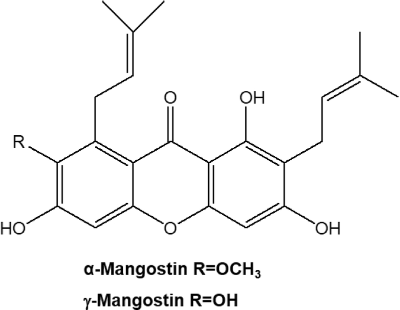
Chemical structures of α- and γ-mangostin isolated from the fruits of Garcinia mangostana.
Evaluation of cell growth inhibition – The inhibitory effect of α- and γ-mangostin on cell proliferation was determined using the sulforhodamine B (SRB) assay.24 Briefly, A549 and resistant A549 cell lines (1 × 10⁴ cells in 190 μL of complete RPMI1640) were seeded in a 96-well plate with various concentrations of mangostins and incubated for 72 h. Then cells were fixed with 10% TCA solution and stained with 0.4% SRB in 1% acetic acid solution. Stained cells were dissolved in 10 mM Tris buffer (pH 10.0), and cell growth was expressed as a percentage relative to the vehicle-treated control. Dose–response curves were analyzed by four-parameter logistic nonlinear regression with a variable slope using GraphPad Prism (GraphPad Software, San Diego, CA, USA). Three independent experiments (n = 3) were performed, and a dose–response curve was fitted separately for each experiment. For each fitted curve, the concentration corresponding to 50% cell viability relative to the vehicle-treated control was determined by interpolation and defined as the IC50. The reported IC50 values represent the mean ± SD of the three independent experiments. The same experimental design and curve-fitting procedure were applied to both parental A549 cells and the resistant cell lines.
Real-time monitoring of cell growth inhibition – Real-time and continuous monitoring of cell growth inhibition was performed by using the xCELLigence system (Roche Applied Science and ACEA Biosciences). Cells (1 × 10⁴ cells/well) were plated in E-plate 96. After 24 h, the cells were treated with various concentrations of mangostins and subsequently monitored every 10 min for a period of up to 72 h by the xCELLigence system. xCELLigence cell index (CI) impedance measurement was performed according to the manufacturer’s instructions and as previously described in detail.25
Western blot analysis – Following treatment with the indicated concentrations of α- or γ-mangostin, whole-cell lysates were prepared for immunoblot analysis. Equal amounts of protein were separated by SDS-PAGE (6–15%) and transferred to PVDF membranes (Millipore, Bedford, MA, USA). The membranes were blocked with 5% skim milk in PBST for 1 h at room temperature and incubated overnight at 4°C with primary antibodies (1:200–1:2000) prepared in 3% skim milk/PBST. After washing with PBST, the appropriate secondary antibodies (1:1000–1:5000) were applied for 2 h at room temperature. Protein bands were detected using an enhanced chemiluminescence (ECL) detection kit (LabFrontier, Suwon, Korea) and LAS-3000 imaging system (Fujifilm, Tokyo, Japan).
Statistical analysis – Data were presented as means ± SD for the indicated number of independently performed experiments. Statistical significance (p < 0.05) was assessed by one-way analysis of variance (ANOVA) coupled with the Dunnett’s t-test.
Results and Discussion
Naturally occurring xanthones have exhibited notable anti-proliferative activities against various human cancer cells.26 Based on these observations, the effects of α- and γ-mangostin on the proliferation of human lung cancer cells were evaluated. As shown in Fig. 2, α- and γ-mangostin inhibited A549 cell proliferation in a concen-tration-dependent manner with the IC50 values of 1.6 and 0.7 μM, respectively (Table 1). The IC50 value of ellipticine, used as a positive control, was 0.5 μM in the A549 cells under the same experimental conditions. In addition, the real-time growth inhibitory effects of α- and γ-mangostin were confirmed using a microelectronic cell sensor system (Fig. 3). The A549 cells were treated with various concentrations of α- and γ-mangostin (0−20 μM), and their growth was monitored. Treatment with 4 or 20 μM α- or γ-mangostin caused the cell index to decrease below its initial value (0 h). In particular, the cell index decreased dramatically to baseline following treatment with 20 μM. These results indicate that α- and γ-mangostin exert cytotoxic effects at higher concentrations (> 4 μM). These findings demonstrate that both mangostins effectively suppress the proliferation of A549 cells, with γ-mangostin exhibiting greater potency than α-mangostin. While α-mangostin has been extensively investigated in NSCLC models,19–21 relatively little information is available regarding γ-mangostin in lung cancer. The lower IC50 value of γ-mangostin observed in the present study suggests that this less-studied xanthone may possess particularly strong anti-proliferative activity and therefore warrants further investigation for its therapeutic potential in NSCLC. However, a limitation of the present study is that the effects of α- and γ-mangostin were evaluated only in NSCLC cell lines, without comparison to nonmalignant human lung cells. Therefore, further studies are needed to evaluate the cancer-cell selectivity and safety profile of these compounds.
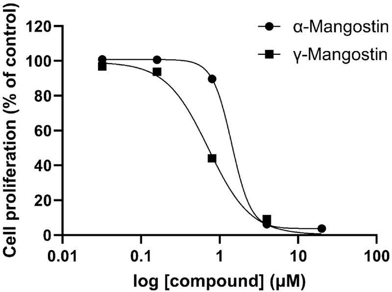
Anti-proliferative effect of α- and γ-mangostin in A549 human lung cancer cells. Cells were treated with the indicated concentrations of α- and γ-mangostin for 72 h, and cell growth was determined by the SRB assay. Dose–response curves were fitted using four-parameter logistic nonlinear regression with a variable slope. A representative curve from three independent experiments is shown.

Inhibitory effect of α- and γ-mangostin on the proliferation of parental and drug-resistant A549 Cells.
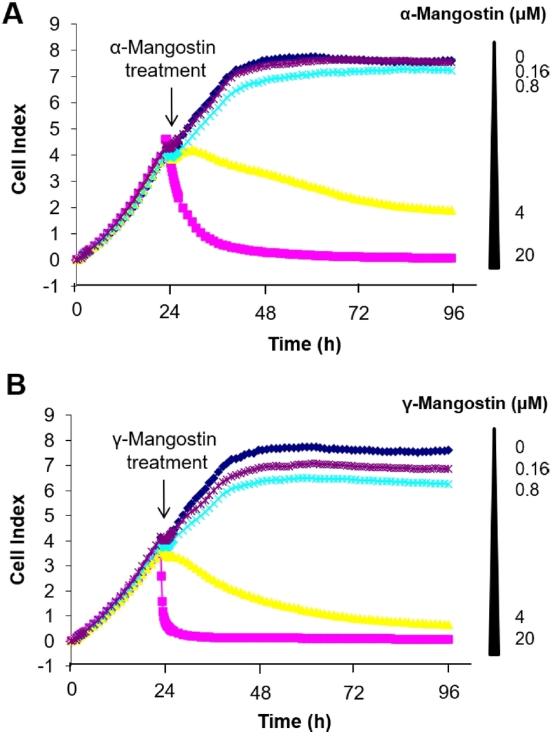
Real-time cellular growth inhibitory activities of α- and γ-mangostin. Cell index curves for cells treated with vehicle control (dark blue), 0.16 μM (purple), 0.8 μM (blue), 4 μM (yellow), or 20 μM (pink) α- and γ-mangostin were measured by the xCELLigence System. Cell index is a measurement derived from electrical impedance of cells interacting with a microelectrode on the bottom of the 96-well plate. Curves represent mean values of three independent experiments.
For patients with newly diagnosed stage IV, relapsed, or recurrent lung cancer, combination chemotherapy consisting of platinum agents, paclitaxel, and gemcitabine remains an important treatment option.27 However, chemotherapy resistance is still an issue in the treatment of lung cancer.28 To the best of our knowledge, the anti-proliferative activities of α- and γ-mangostin in drug-resistant lung cancer cells have not previously been reported. Therefore, we investigated whether α- and γ-mangostin could overcome resistance to conventional anticancer drugs, such as paclitaxel and gemcitabine, in A549 cells. Paclitaxel- and gemcitabine-resistant A549 cell lines (A549-Pac and A549-Gem, respectively) were established by repeated exposure to stepwise increasing concentrations of each drug for more than six months. A549-Pac and A549-Gem cells were 188- and 326-fold more resistant to paclitaxel and gemcitabine, respectively, than parental cells (Table 1). α- and γ-mangostin, however, inhibited the growth of parental, A549-Pac, and A549-Gem cells to a similar extent, suggesting that these compounds retain their anti-proliferative activity in paclitaxel- and gemcitabine-resistant cells. Although the molecular basis underlying this effect remains to be elucidated, the ability of both compounds to overcome chemo-resistance supports their therapeutic potential in drug-resistant NSCLC. Notably, γ-mangostin retained strong activity across both parental and resistant cell lines, further highlighting its therapeutic potential.
Given that KRAS mutations constitutively activate multiple downstream signaling pathways, including ERK, PI3K/AKT, and STAT3, which are closely associated with tumor progression and therapeutic resistance, we investigated whether α-mangostin and γ-mangostin exert their anticancer effects through modulation of these KRAS-associated signaling networks in A549 lung cancer cells. To evaluate the effects of α- and γ-mangostin on KRAS-associated downstream signaling, we first examined the PI3K/AKT/mTOR pathway. As shown in Fig. 4A, treatment with α- and γ-mangostin markedly reduced the phosphorylation levels of Akt and mTOR. Consistent with the inhibition of mTOR signaling, the phosphorylation of its downstream effectors, eukaryotic translation initiation factor 4E-binding protein 1 (4E-BP1) and p70 ribosomal protein S6 kinase (p70S6K), was also significantly decreased. Since AMP-activated protein kinase (AMPK) is a critical negative regulator of mTOR signaling, we further assessed AMPK activation and found that both mangostins increased the phosphorylation of AMPK compared to the vehicle-treated control group. These findings suggest that α- and γ-mangostin suppress the KRAS-associated PI3K/AKT/mTOR signaling axis, as evidenced by reduced phosphorylation of Akt, mTOR, and their downstream effectors. The observed activation of AMPK may additionally contribute to mTOR inhibition, indicating that mangostins regulate mTOR signaling through multiple upstream mechanisms.
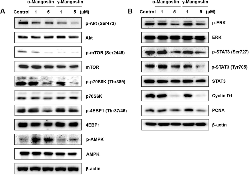
Effects on the expression of signal transduction proteins. A549 cells were treated with α- and γ-mangostin (1 or 5 μM) for 24 h, and the protein expression was measured by Western blot analysis. Representative blots from three independent experiments are shown.
We next investigated the effects of α- and γ-mangostin on the MAPK/ERK and STAT3 pathways, two additional major associated downstream effectors of oncogenic KRAS signaling. As shown in Fig. 4B, both compounds significantly inhibited ERK phosphorylation. Notably, STAT3 activation, as determined by phosphorylation at Ser727 and Tyr705 residues, was also markedly suppressed by mangostin treatment. Taken together, these findings indicate that α- and γ-mangostin suppress both ERK and STAT3 signaling, further supporting their ability to inhibit multiple KRAS-associated downstream pathways in A549 lung cancer cells.
To determine whether the inhibition of KRAS-associated downstream signaling translated into suppression of cell proliferative capacity, we further analyzed the expression of proliferation-related biomarkers. As shown in Fig. 4B, α- and γ-mangostin strongly suppressed cyclin D1 expression and moderately reduced PCNA levels. Collectively, these findings indicate that α- and γ-mangostin inhibit multiple KRAS-associated downstream signaling pathways, including PI3K/AKT/mTOR, ERK, and STAT3, leading to the suppression of proliferation-associated proteins and contributing to their anti-pro-liferative effects in A549 lung cancer cells.
Since KRAS-mutant NSCLC is characterized by persistent activation of downstream survival signaling and is frequently associated with resistance to EGFR-targeted therapies, we further investigated whether α- and γ-mangostin could suppress EGF-induced Akt activation in A549 cells. As shown in Fig. 5, serum-starved A549 cells exhibited low basal levels of phosphorylated Akt, whereas stimulation with EGF (10 ng/mL) for 10 min markedly increased Akt phosphorylation. However, treatment with α- and γ-mangostin (1 μM) significantly attenuated EGF-induced Akt activation. The ability of α- and γ-mangostin to suppress Akt activation under both basal and EGF-stimulated conditions suggests that these compounds can modulate survival signaling induced by extracellular growth factors as well as constitutively activated pathways. Although direct effects on resistance to EGFR-targeted therapies were not evaluated in this study, attenuation of EGF-induced Akt activation may be relevant to signaling mechanisms underlying therapeutic resistance in NSCLC.10–12 Further studies using models of EGFR-targeted therapy-resistant NSCLC will be necessary to determine the relevance of these findings in resistant tumors.
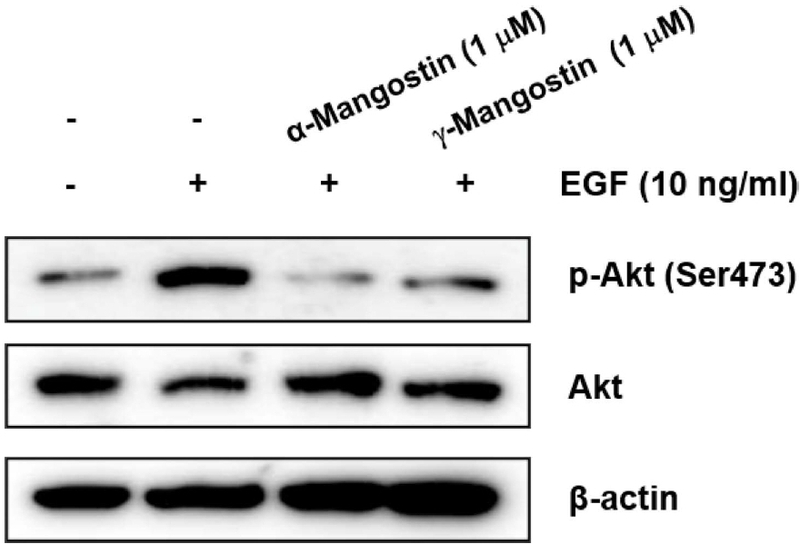
Effects on EGF-stimulated Akt signaling by α- and γ-mangostin. A549 cells were serum starved for 24 h and then treated with α- and γ-mangostin (1 μM) for 2 h, followed by EGF (10 ng/mL) stimulation for 10 min. The expression of Akt was determined by Western blots. Representative blots from three independent experiments are shown.
In summary, α- and γ-mangostin exhibited potent anti-proliferative activity against both parental and chemo-resistant human lung cancer cells. Their anti-proliferative effects were associated with inhibition of PI3K/AKT/mTOR, ERK, and STAT3 signaling pathways. Collectively, these findings suggest that α- and γ-mangostin act as multi-target modulators of oncogenic signaling networks and may have therapeutic potential for KRAS-mutant and drug-resistant NSCLC. In particular, the robust activity of γ-mangostin observed in this study highlights its potential as a relatively underexplored compound for future lung cancer research.
Acknowledgments
This work was supported by the National Research Foundation of Korea (NRF) grant funded by the Korea government (MSIT) (RS-2025-23963632).
Conflicts of Interest
The authors declare that they have no conflicts of interest.
References
-
Siegel, R. L.; Kratzer, T. B.; Wagle, N. S.; Sung, H.; Jemal, A. CA Cancer J. Clin. 2026, 76, e70043.
[https://doi.org/10.3322/caac.70043]

-
Gridelli, C.; Rossi, A.; Carbone, D. P.; Guarize, J.; Karachaliou, N.; Mok, T.; Petrella, F.; Spaggiari, L.; Rosell, R. Nat. Rev. Dis. Primers 2015, 1, 15009.
[https://doi.org/10.1038/nrdp.2015.9]
-
Ashrafi, A.; Akter, Z.; Modareszadeh, P.; Modareszadeh, P.; Berisha, E.; Alemi, P. S.; Chacon Castro, M. D. C.; Deese, A. R.; Zhang, L. Cancers (Basel) 2022, 14, 4562.
[https://doi.org/10.3390/cancers14194562]
-
Westcott, P. M.; To, M. D. Chin. J. Cancer 2013, 32, 63–70.
[https://doi.org/10.5732/cjc.012.10098]
-
Tang, Y.; Pu, X.; Yuan, X.; Pang, Z.; Li, F.; Wang, X. Cancer Gene Ther. 2024, 31, 961–969.
[https://doi.org/10.1038/s41417-024-00778-4]
-
Asati, V.; Mahapatra, D. K.; Bharti, S. K. Eur. J. Med. Chem. 2016, 109, 314–341.
[https://doi.org/10.1016/j.ejmech.2016.01.012]
-
Wang, W.; Alley, A.; Sun, N.; Tadesse, M.; Wang, X.; Zhang, R. Front. Pharmacol. 2025, 16, 1663766.
[https://doi.org/10.3389/fphar.2025.1663766]
-
Gough, D. J.; Koetz, L.; Levy, D. E. PLoS One 2013, 8, e83395.
[https://doi.org/10.1371/journal.pone.0083395]
-
Yoon, Y. K.; Kim, H. P.; Han, S. W.; Oh, D. Y.; Im, S. A.; Bang, Y. J.; Kim, T. Y. Mol. Carcinog. 2010, 49, 353–362.
[https://doi.org/10.1002/mc.20607]
-
Pao, W.; Wang, T. Y.; Riely, G. J.; Miller, V. A.; Pan, Q.; Ladanyi, M.; Zakowski, M. F.; Heelan, R. T.; Kris, M. G.; Varmus, H. E. PLoS Med. 2005, 2, e17.
[https://doi.org/10.1371/journal.pmed.0020017]
-
Mao, C.; Qiu, L. X.; Liao, R. Y.; Du, F. B.; Ding, H.; Yang, W. C.; Li, J.; Chen, Q. Lung Cancer 2010, 69, 272–278.
[https://doi.org/10.1016/j.lungcan.2009.11.020]
-
Shin, D. H.; Jo, J. Y.; Choi, M.; Kim, K. H.; Bae, Y. K.; Kim, S. S. Exp. Mol. Med. 2023, 55, 2220–2237.
[https://doi.org/10.1038/s12276-023-01091-0]
-
Dehelean, C. A.; Marcovici, I.; Soica, C.; Mioc, M.; Coricovac, D.; Iurciuc, S.; Cretu, O. M.; Pinzaru, I. Molecules 2021, 26, 1109.
[https://doi.org/10.3390/molecules26041109]
-
Jung, H. A.; Su, B. N.; Keller, W. J.; Mehta, R. G.; Kinghorn, A. D. J. Agric. Food Chem. 2006, 54, 2077–2082.
[https://doi.org/10.1021/jf052649z]
-
Pedraza-Chaverri, J.; Cardenas-Rodriguez, N.; Orozco-Ibarra, M.; Perez-Rojas, J. M. Food Chem. Toxicol. 2008, 46, 3227–3239.
[https://doi.org/10.1016/j.fct.2008.07.024]
-
Obolskiy, D.; Pischel, I.; Siriwatanametanon, N.; Heinrich, M. Phytother. Res. 2009, 23, 1047–1065.
[https://doi.org/10.1002/ptr.2730]
-
Majdalawieh, A. F.; Terro, T. M.; Ahari, S. H.; Abu-Yousef, I. A. Biomolecules 2024, 14, 1382.
[https://doi.org/10.3390/biom14111382]
-
Chang, H. F.; Yang, L. L. Molecules 2012, 17, 8010–8021.
[https://doi.org/10.3390/molecules17078010]
-
Zhang, C.; Yu, G.; Shen, Y. Saudi J. Biol. Sci. 2018, 25, 1090–1095.
[https://doi.org/10.1016/j.sjbs.2017.03.005]
-
Ding, Y. Y.; Luan, J. J.; Fan, Y.; Olatunji, O. J.; Song, J.; Zuo, J. Cell Stress Chaperones 2020, 25, 163–172.
[https://doi.org/10.1007/s12192-019-01063-2]
-
Shih, Y. W.; Chien, S. T.; Chen, P. S.; Lee, J. H.; Wu, S. H.; Yin, L. T. Cell Biochem. Biophys. 2010, 58, 31–44.
[https://doi.org/10.1007/s12013-010-9091-2]
-
Sarmoko, S.; Novitasari, D.; Toriyama, M.; Fareza, M. S.; Choironi, N. A.; Itoh, H.; Meiyanto, E. Iran. J. Pharm. Res. 2023, 22, e138856.
[https://doi.org/10.5812/ijpr-138856]
-
Quan, G. H.; Oh, S. R.; Kim, J. H.; Lee, H. K.; Kinghorn, A. D.; Chin, Y. W. Phytother. Res. 2010, 24, 1575–1577.
[https://doi.org/10.1002/ptr.3177]
-
Song, S. H.; Min, H. Y.; Lee, S. K. Biomol. Therap. 2010, 18, 402–410.
[https://doi.org/10.4062/biomolther.2010.18.4.402]
-
Solly, K.; Wang, X.; Xu, X.; Strulovici, B.; Zheng, W. Assay Drug Dev. Technol. 2004, 2, 363–372.
[https://doi.org/10.1089/adt.2004.2.363]
-
Pedro, M.; Cerqueira, F.; Sousa, M. E.; Nascimento, M. S.; Pinto, M. Bioorg. Med. Chem. 2002, 10, 3725–3730.
[https://doi.org/10.1016/S0968-0896(02)00379-6]
- PDQ® Adult Treatment Editorial Board. PDQ Non-Small Cell Lung Cancer Treatment. Bethesda, MD: National Cancer Institute. Updated <05/15/2025>. Available at: https://www.cancer.gov/types/lung/hp/non-small-cell-lung-treatment-pdq, . Accessed <06/17/2026>. [PMID: 26389304].
-
Cao, Z.; Zhu, J.; Chen, X.; Chen, Z.; Wang, W.; Zhou, Y.; Hua, Y.; Shi, J.; Chen, J. J. Thorac. Dis. 2024, 16, 8039–8052.
[https://doi.org/10.21037/jtd-24-1078]