
Lonicerin from Lonicera japonica Attenuates Scopolamine-Induced Memory Impairment in Mice
Abstract
This study examined whether lonicerin, a flavonoid glycoside isolated from Lonicera japonica, could reverse memory deficits induced by scopolamine administration in mice, drawing on both behavioral testing and biochemical analysis. Spatial learning and memory were evaluated using the Morris water maze, where animals given lonicerin displayed markedly shorter escape latencies, reduced path lengths, and more efficient swimming trajectories relative to scopolamine-only controls. A separate passive avoidance paradigm confirmed that lonicerin restored retention memory to roughly 65% of the level observed in untreated control animals. Biochemically, lonicerin treatment was associated with a significant suppression of acetylcholinesterase activity in the hippocampus, a region central to memory formation. Together, these findings point to acetylcholinesterase inhibition as a key mechanism through which lonicerin counteracts scopolamine-induced cholinergic dysfunction, supporting its further evaluation as a candidate agent for memory impairment and neurodegenerative disease.
Keywords:
Lonicera japonica, Lonicerin, Alzheimer's disease, Cognitive-enhancing activity, AcetylcholinesteraseIntroduction
Alzheimer's disease is a progressive, age-related neurodegenerative condition most often diagnosed after the age of 65, characterized pathologically by the accumulation of amyloid plaques and tau tangles alongside chronic neuroinflammation and gradual neuronal loss. Clinically, the disease typically presents first as memory impairment before advancing to broader cognitive decline. Central to this process is dysfunction of the cholinergic system, since acetylcholine-dependent signaling underlies much of learning and memory function.1 As cholinergic neurons are lost and acetylcholine availability falls, memory and cognitive performance deteriorate accordingly, a relation ship that has made the cholinergic system a primary target for therapeutic intervention.2 Loss of cholinergic neurons within the basal forebrain is regarded as one of the pathological hallmarks of Alzheimer's disease, and pharmacological agents that inhibit acetylcholinesterase — including physostigmine, tacrine, and donepezil — have been shown in behavioral studies to alleviate spatial memory deficits.3-5
Blockade of central muscarinic receptors by scopolamine produces a reversible but pronounced disruption of cholinergic signaling, and the resulting deficits — impaired attention, slowed information processing, and difficulty forming new memories — are observed in both animal models and human subjects, closely paralleling the cognitive symptoms of Alzheimer's disease.6-9 For this reason, scopolamine-induced amnesia in mice has become a standard experimental paradigm for probing the mechanisms underlying Alzheimer's-type cognitive decline and for screening candidate therapeutics.10,11
In earlier work from our laboratory, we characterized the neuroprotective activity of lonicerin, a bioactive constituent of Lonicera japonica, using the HT-22 hippo-campal neuronal cell line as a model of glutamate-induced excitotoxicity. That study found that lonicerin attenuated intracellular calcium (Ca2+) overload and peroxide accumulation triggered by glutamate exposure, while also helping to preserve glutathione (GSH) levels, thereby limiting oxidative injury in cultured cortical neurons.12 Also, we demonstrated that the crude extract of L. japonica effectively ameliorates scopolamine-induced memory deficits through cholinergic modulation.13 As a critical follow-up to identify the active single constituents responsible for these bioactivities, the present study evaluated the in vivo cognitive-enhancing effects of lonicerin, a major flavone glycoside isolated from L. japonica. By validating the therapeutic efficacy of isolated lonicerin in a mouse model of cognitive impairment, this study provides distinct novel insights into the neuroprotective mechanisms of specific phytochemicals derived from L. japonica.
Building on those findings, the present study asked whether lonicerin could also reverse cognitive impairment in vivo, specifically in mice rendered amnesic by scopolamine. Learning and memory were assessed using two established behavioral paradigms — the passive avoidance test and the Morris water maze — in order to gauge the compound's therapeutic potential against memory dysfunction.
To probe the mechanism behind any observed behavioral effects, acetylcholinesterase activity was measured in the hippocampus of scopolamine-treated animals, since inhibition of this enzyme is expected to enhance cholinergic transmission and, in turn, cognitive performance in states of cholinergic deficiency such as Alzheimer's disease. Overall, this work sought to characterize the cognitive-enhancing potential of lonicerin and its possible relevance as a therapeutic candidate for memory impairment, with an eye toward informing future strategies for treating neurodegenerative disease and other disorders marked by cognitive decline.
Experimental
Experimental animals – Male ICR mice (4 weeks old, approximately 25 g body weight) were used to evaluate cognitive-enhancing activity. Animals were supplied by Kangwon Life Science Co. (Gangwon Province, Korea) and housed at the Animal Care Center of Kangwon National University. Following a one-week acclimatization period, mice were maintained under controlled conditions (23 ± 1°C, 60% relative humidity, 12-hour light/dark cycle) with free access to food and water. All experimental procedures adhered to the Kangwon National University Animal Care and Use Guidelines (IACUC approval no. KIACUC, 08/2024) and were conducted in accordance with the ARRIVE guidelines for animal research. All animal cohorts, control groups (vehicle and donepezil), and behavioral/biochemical datasets in this study were generated completely independently from our previous investigation on L. japonica extract study. No animals, tissues, or raw data were shared or reused between the extract study and the present lonicerin study.
Compounds or drugs – Lonicerin (purity >95%, HPLC) was isolated and purified from L. japonica in according to the previously established extraction and chromatographic purification protocol.14 To secure a sufficient quantity ( ≥ 12.0 mg) for all in vivo behavioral and biochemical evaluations, the identical isolation and purification process was repeated across multiple batches. Scopolamine (purity >98%) was purchased from Sigma (St. Louis, MO, USA), together with phosphate-buffered saline (PBS) and carboxymethyl cellulose (CMC); it was used to induce cognitive deficits by elevating acetylcholinesterase activity, mimicking key features of Alzheimer's disease pathology. Donepezil (purity >95%), obtained from Samjin Pharmaceutical Co., Ltd. (Seoul, Korea), served as the positive-control reference compound. Scopolamine is well established as an inducer of Alzheimer's-like amnesia, acting by depleting brain acetylcholine through heightened acetylcholinesterase activity and thereby impairing memory and cognition. Donepezil, by contrast, acts by inhibiting acetylcholine-sterase and so prevents acetylcholine breakdown, sustaining elevated acetylcholine levels that support memory retention and cognitive performance; owing to this efficacy, it is widely used clinically for Alzheimer's disease and commonly serves as the benchmark comparator in studies of candidate anti-dementia agents.15
Drug administration – Mice were divided into six experimental groups of six animals each: an untreated control group, a scopolamine-only group, a positive-control group receiving donepezil (1 mg/kg), and three groups receiving lonicerin at 1, 5, or 10 mg/kg. Dose selection was guided by prior in vitro findings and by considerations relevant to potential clinical application. No mortality or overt signs of toxicity occurred during the study, and post-experiment organ examination revealed no abnormalities relative to controls. Lonicerin and donepezil were administered orally 90 minutes prior to scopolamine injection, while control animals received 0.5% carboxymethyl cellulose (CMC) vehicle instead. Scopolamine (1 mg/kg) was injected subcutaneously 30 minutes before behavioral testing began. Dosing continued for four consecutive days, with an additional dose given on the training day of the passive avoidance test. Cognitive performance was assessed using both the Morris water maze and passive avoidance paradigms.
Morris water maze test – Spatial memory was evaluated using a modified version of the Morris water maze procedure.16 Testing was conducted in a circular pool (90 cm diameter, 40 cm depth) filled with water held at 20 ± 1°C; 500 mL of white milk was added to render the submerged escape platform invisible. The pool was conceptually divided into four quadrants, with a platform (10 cm diameter, 26 cm height) submerged 1 cm below the surface in one quadrant. Swimming behavior was tracked with a camera linked to a video-tracking system (Smart ver. 2.5.21) that recorded latency, distance traveled, and swim speed, with escape latency — the time required for the animal to locate and mount the platform — serving as the primary outcome measure. On day one, mice were given a 60-second habituation swim without the platform present. Over the following four days, each mouse received up to 120 seconds per trial to locate the platform; if the platform was not found within this window, a ceiling latency of 120 seconds was recorded.
Passive avoidance test – The passive avoidance test was conducted using the same cohort of mice that had previously completed the Morris water maze test. The passive avoidance procedure, adapted from our previous protocol, employed a two-compartment apparatus (17 × 12 × 10 cm) divided by a guillotine door, with the dark compartment fitted with an electrified grid floor. Testing proceeded across three stages – acquisition, training, and test. During acquisition, mice were placed in the lit compartment and permitted 40 seconds of free exploration before the guillotine door opened; the door was closed once the animal entered the dark compartment. Twenty-four hours later, during the training stage, mice were again placed in the lit compartment; after a 30-second delay the door opened, and upon entry into the dark compartment the animal received a brief foot shock (0.1 mA/10 g body weight, 2 s). The test stage, conducted 24 hours after training, measured the latency to enter the dark compartment (maximum 180 seconds) as the primary index of retention memory. Mice were euthanized by cervical dislocation upon completion of testing.
Acetylcholinesterase (AChE) inhibition assay – Hippocampal tissues from all six mice in each group (n = 6) were used to evaluate acetylcholinesterase (AChE) activity. For each individual sample, the assay was performed in triplicates to ensure measurement accuracy, and the mean value of the triplicates was taken as the representative value for that animal. Acetylcholinesterase activity was measured using a modified Ellman assay to determine the effect of lonicerin treatment. Brain tissue was harvested within 30 minutes of completing behavioral testing (Morris water maze or passive avoidance), and hippocampal tissue was dissected and homogenized in ice-cold sodium phosphate buffer before centrifugation to obtain a clear supernatant. Assay mixtures contained hippocampal supernatant, sodium phosphate buffer, 5,5′-dithiobis(2-nitrobenzoic acid) (DTNB), and acetylcholine iodide (AChI); the reaction was initiated by addition of AChI, and thiocholine production was monitored spectrophotometrically at 412 nm using a microplate reader. All assays were run in triplicate alongside non-enzymatic blanks.
Statistics – Data from the Morris water maze, passive avoidance test, and AChE assay are expressed as mean ± standard error of the mean (S.E.M.). Group differences were evaluated by one-way ANOVA followed by Tukey's post hoc test, using IBM SPSS Statistics (version 26). Statistical significance was set at p < 0.05, p < 0.01, and p < 0.001.
Results and Discussion
The Morris water maze was used to examine whether lonicerin could counteract scopolamine-induced spatial memory deficits (Fig. 1). Escape latency in the control group fell steadily across the four testing days, reaching 40.5 seconds by the final day, whereas the scopolamine-only group showed a prolonged and largely unchanging latency that peaked at 109.4 seconds on day four — confirming that scopolamine effectively impaired spatial memory. Improvement in the treatment groups became apparent from day three onward. The donepezil group recorded a latency of 65.1 seconds on day three, which fell further to approximately 54.6 seconds by day four. Lonicerin-treated animals likewise showed a dose-dependent reduction in escape latency relative to the scopolamine group: latencies of 67.2, 84.1, and 95.3 seconds were recorded at the 10, 5, and 1 mg/kg doses, respectively (Fig. 2).
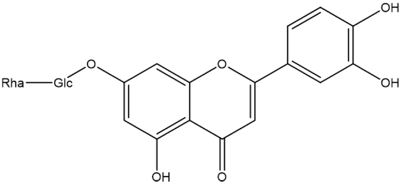
Chemical structure of lonicerin isolated from L. japonica.
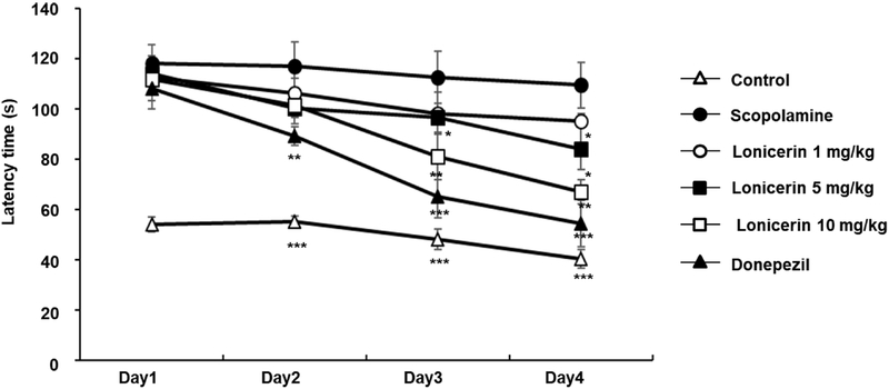
Effects of lonicerin on spatial learning and memory in the Morris water maze test. Escape latency is presented as the mean ± SD (n = 6). *p < 0.05, **p < 0.01, ***p < 0.001 compared with the scopolamine-treated group.
A similar pattern emerged in total swimming distance. The control group covered a significantly shorter distance (320 cm) than the scopolamine-treated group (654 cm), roughly double, reflecting the pronounced impairment of spatial learning and memory induced by scopolamine. Across the four-day testing period, lonicerin administration reduced this excess swimming distance in a dose-dependent manner: mice given 1 mg/kg covered 633 cm, a moderate improvement over the scopolamine group, while those given 5 mg/kg and 10 mg/kg covered 548 cm and 501 cm, respectively — a clear trend toward better spatial performance with increasing dose (Fig. 3).
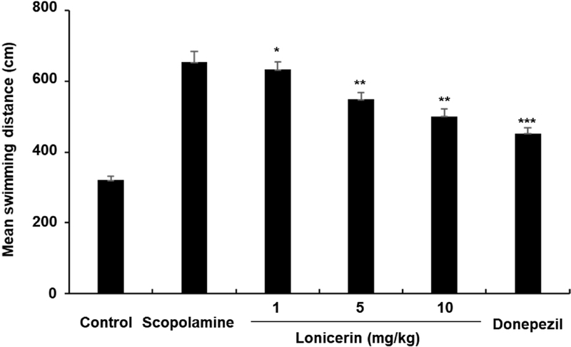
Effects of lonicerin on swimming distance in the Morris water maze test. Data are expressed as the mean ± SD (n = 6). *p < 0.05, **p < 0.01, ***p < 0.001 compared with the scopolamine-treated group.
Memory retention was further assessed with a probe trial measuring the time spent in the target quadrant that had previously contained the escape platform. Control animals spent considerably longer in this quadrant (39.2 seconds) than scopolamine-treated animals (9.4 seconds), underscoring both the intact retention of the control group and the memory-impairing effect of scopolamine. Lonicerin treatment increased target-quadrant dwell time in a dose-dependent fashion, with values of 12.1, 30.4, and 30.7 seconds at the 1, 5, and 10 mg/kg doses, respectively — indicating that higher doses more effectively reversed the scopolamine-induced deficit in memory retention (Fig. 4).
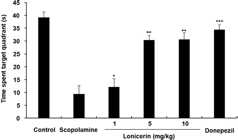
Effects of lonicerin on spatial memory retention in the probe trial of the Morris water maze. Data are presented as the mean ± SD (n = 6). *p < 0.05, **p < 0.01, ***p < 0.001 compared with the scopolamine-treated group.
Because faster platform discovery could in principle reflect enhanced motor performance rather than improved memory, swimming speed was also compared across groups. No significant differences in mean swim speed were found among the control, scopolamine, donepezil, and lonicerin-treated groups (Fig. 5), indicating that the shorter escape latencies and increased quadrant dwell times seen with lonicerin and donepezil reflect genuine improvements in spatial memory rather than changes in locomotor capacity. This consistency in swim speed across groups helps isolate the memory-enhancing effects of the treatments from any confounding influence of physical performance.
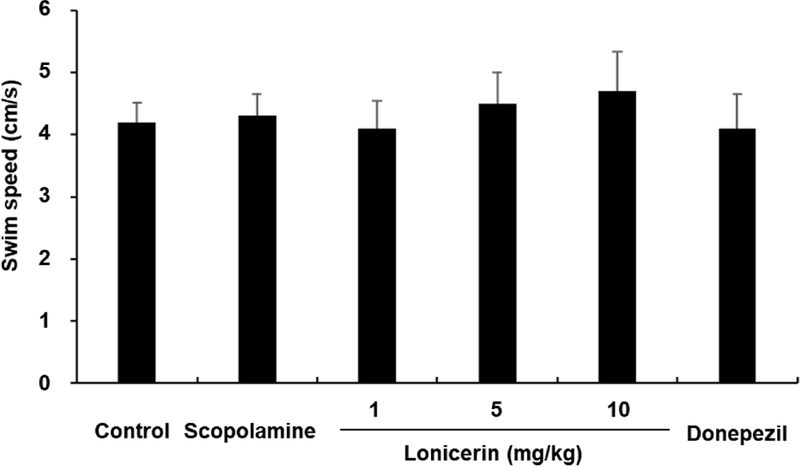
Effects of lonicerin on swimming speed during the Morris water maze test. Data are expressed as the mean ± SD (n = 6). *p < 0.05, **p < 0.01, ***p < 0.001 compared with the scopolamine-treated group.
Scopolamine disrupts cholinergic signaling and is known to raise hippocampal AChE activity, producing the learning and memory deficits captured by escape latency in the water maze.17 Lonicerin shortened escape latency in a dose-dependent manner, with higher doses yielding faster platform discovery relative to scopolamine-treated mice, while swim speed remained comparable across the control, scopolamine, and lonicerin groups throughout the four-day test — again pointing to a memory-specific rather than motor effect. Mice treated with lonicerin also spent more time in the target quadrant than scopolamine-treated animals, consistent with improved recall of the platform location. Taken together, these results indicate that lonicerin improves spatial learning and memory independently of any change in swimming ability.
The passive avoidance test, a well-established measure of long-term memory, was used to determine whether the benefits of lonicerin extended beyond the water maze paradigm to a fear-motivated learning task. In this test, mice learn to avoid a compartment paired with an aversive stimulus, and the latency to re-enter that compartment provides a measure of retained information over time.18 During the acquisition trial, latency to enter the dark compartment did not differ meaningfully across groups, indicating comparable initial learning ability: mean latencies were 2.11 s (control), 2.32 s (scopolamine), and 2.31 s (donepezil), with the lonicerin groups recording 2.18 s (1 mg/kg), 2.44 s (5 mg/kg), and 2.13 s (10 mg/kg) (Fig. 6).
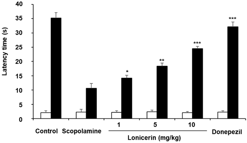
Effects of lonicerin on long-term memory performance in the passive avoidance test. Step-through latency is expressed as the mean ± SD (n = 6). *p < 0.05, **p < 0.01, ***p < 0.001 compared with the scopolamine-treated group.
In the subsequent test trial, the scopolamine group showed a markedly shorter latency than the control group (10.58 s versus 35.22 s), confirming that scopolamine produced the expected memory impairment. Lonicerin-treated groups, by contrast, showed dose-dependent increases in latency — 14.22 s, 18.32 s, and 24.51 s at 1, 5, and 10 mg/kg, respectively (Fig. 6). Given that acquisition-phase learning was equivalent across groups, this dose-dependent rise in test-trial latency indicates that lonicerin specifically enhanced retention memory and partially reversed the amnesic effect of scopolamine.
Hippocampal AChE activity was elevated by 158.2% in the scopolamine group relative to control, consistent with scopolamine's known effect of increasing enzymatic breakdown of acetylcholine. Lonicerin treatment significantly reduced this elevation in a dose-dependent manner (p < 0.05): AChE activity was reduced by 132.5%, 125.2%, and 113.7% at the 1, 5, and 10 mg/kg doses, respectively, relative to the scopolamine group (Fig. 7).
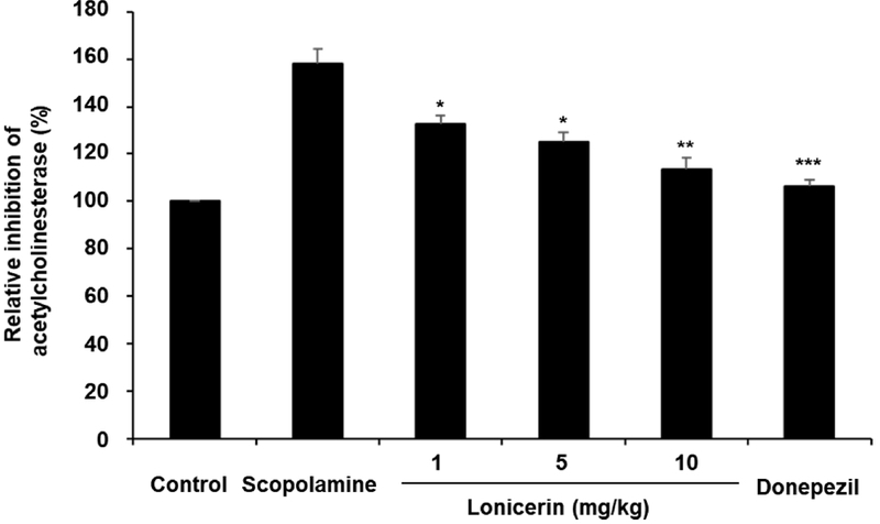
Inhibitory effect of lonicerin on acetylcholinesterase (AChE) activity. Data are presented as the mean ± SD (n = 6 per group; each sample was assayed in triplicates). *p < 0.05, **p < 0.01, ***p < 0.001 compared with the scopolamine-treated group.
Because acetylcholine availability is central to learning and memory, and because reduced acetylcholine levels in Alzheimer's disease are closely tied to elevated AChE activity, this enzyme remains a principal pharmacological target; suppressing AChE activity helps preserve acetylcholine and, in turn, supports cognitive function.19,20 Established AChE inhibitors such as donepezil, galantamine, and tacrine act through this same mechanism, prolonging acetylcholine availability and thereby improving cholinergic transmission and cognitive performance in conditions of acetylcholine deficiency.20,21 In the present study, lonicerin showed dose-dependent AChE-inhibitory activity, with higher doses producing a greater reduction in enzyme activity, consistent with a mechanism in which lonicerin helps sustain brain acetylcholine levels and thereby alleviates scopolamine-induced memory deficits. That said, the AChE-inhibitory potency of lonicerin observed here is modest rather than pronounced.
Lonicerin is a flavone glycoside composed of luteolin conjugated with a rutinoside moiety. As a member of the flavonoid family, it possesses structural features associated with antioxidant and anti-inflammatory activities, both of which are increasingly recognized as important therapeutic strategies for neurodegenerative disorders.22,23 Previous studies have demonstrated that lonicerin exhibits diverse pharmacological activities, including antioxidant, anti-inflammatory, antiviral, and hepatoprotective effects. These biological properties are particularly relevant to Alzheimer's disease, where oxidative stress, neuroinflammation, and cholinergic dysfunction act synergistically to drive progressive neuronal injury.24,25 The relatively modest AChE inhibition observed in the present study suggests that cholinergic modulation is unlikely to fully account for the cognitive benefits of lonicerin. Rather, its previously reported antioxidant and neuroprotective properties may act in concert with AChE inhibition, resulting in a multimodal mechanism of action.26
A potential limitation of the present study lies in the inherent methodological characteristic of the scopolamine-induced memory deficit model. Because both the Morris water maze and passive avoidance tests heavily rely on visual cues, scopolamine-induced peripheral cholinergic blockade – such as mydriasis and blurred vision – could potentially confound behavioral outcomes independently of central memory deficits.6 Although swimming speed was monitored to account for motor performance in this study, the inclusion of a non-CNS penetrating anticholinergic control, such as methylscopolamine, would be necessary to fully rule out these peripheral visual artifacts. Future studies incorporating methylscopolamine controls or vision-independent task batteries will be essential to definitively isolate the central neuroprotective effects of lonicerin.
Combined with the neuroprotective activity previously reported for lonicerin by our group, this suggests that its cognitive-enhancing effects likely arise from multiple, complementary mechanisms whose combined impact becomes more apparent with repeated dosing. Future work examining behavioral outcomes and AChE activity after a single administration would help clarify the relative contribution and time-course of these mechanisms.
In summary, lonicerin, isolated from L. japonica, dose-dependently attenuated scopolamine-induced memory impairment across both the Morris water maze and passive avoidance paradigms. This effect appears to be mediated at least in part by inhibition of acetylcholinesterase, the enzyme responsible for degrading the memory-related neurotransmitter acetylcholine; by limiting this degradation, lonicerin helps sustain acetylcholine levels relevant to learning and memory. These findings support the further development of lonicerin as a candidate compound for treating memory impairment associated with neurodegenerative conditions, including Alzheimer's disease.
Acknowledgments
This research was supported by the Korea Basic Science Institute (National Research Facilities and Equipment Center) grant funded by the Ministry of Education (grant No. 2022R1A6C101A739).
Conflicts of Interest
The authors have declared that there are no conflicts of interest.
References
-
Jack, C. R., Jr.; Bennett, D. A.; Blennow, K.; Carrillo, M. C.; Dunn, B.; Haeberlein, S. B.; Holtzman, D. M.; Jagust, W.; Jessen, F.; Karlawish, J.; Liu, E.; Molinuevo, J. L.; Montine, T.; Phelps, C.; Rankin, K. P.; Rowe, C. C.; Scheltens, P.; Siemers, E.; Snyder, H. M.; Sperling, R. Alzheimer's Dement. 2018, 14, 535–562.
[https://doi.org/10.1016/j.jalz.2018.02.018]

-
DeTure, M. A.; Dickson, D. W. Mol. Neurodegener. 2019, 14, 32.
[https://doi.org/10.1186/s13024-019-0333-5]
-
Bartus, R. T.; Dean, R. L., III; Beer, B.; Lippa, A. S. Science 1982, 217, 408–414.
[https://doi.org/10.1126/science.7046051]
-
Giacobini, E. Pharmacol. Res. 2004, 50, 433–440.
[https://doi.org/10.1016/j.phrs.2003.11.017]
- Birks, J. Cochrane Database Syst. Rev. 2006, CD005593.
-
Klinkenberg, I.; Blokland, A. Neurosci. Biobehav. Rev. 2010, 34, 1307–1350.
[https://doi.org/10.1016/j.neubiorev.2010.04.001]
-
Ebert, U.; Kirch, W. Eur. J. Clin. Invest. 1998, 28, 944–949.
[https://doi.org/10.1046/j.1365-2362.1998.00393.x]
-
Bartus, R. T. Exp. Neurol. 2000, 163, 495–529.
[https://doi.org/10.1006/exnr.2000.7397]
-
Pepeu, G.; Giovannini, M. G. Learn. Mem. 2004, 11, 21–27.
[https://doi.org/10.1101/lm.68104]
- Chen, Y.; Sun, J.; Wang, Y.; Lu, S.; Zhu, W.; Wang, X.; Li, X.; Kong, L. Psychopharmacology 2024, 241, 1779–1804.
-
Jagielska, A.; Sałaciak, K.; Pytka, K. Ageing Res. Rev. 2025, 104, 102635.
[https://doi.org/10.1016/j.arr.2024.102635]
- Weon, J. B.; Yang, H. J.; Lee, B.; Yun, B. R.; Ma, C. J. Nat. Prod. Sci. 2011, 17, 221–224.
-
Ma, C. J. Nat. Prod. Sci. 2026, 32, 138–146.
[https://doi.org/10.20307/nps.2026.32.2.138]
-
Lee, H.; Ma, C. J. Kor. J. Pharmacogn. 2021, 52, 19–25.
[https://doi.org/10.36430/kjp.2021.25.2.002]
- Hampel, H.; Mesulam, M. M.; Cuello, A. C.; Farlow, M. R.; Giacobini, E.; Grossberg, G. T.; Khachaturian, A. S.; Vergallo, A.; Cavedo, E.; Snyder, P. J.; Khachaturian, Z. S. Nat. Rev. Neurol. 2018, 14, 377–399.
-
Vorhees, C. V.; Williams, M. T. ILAR J. 2014, 55, 310–332.
[https://doi.org/10.1093/ilar/ilu013]
-
Kim, S. T. Life Sci. 2019, 233, 116695.
[https://doi.org/10.1016/j.lfs.2019.116695]
- Ennaceur, A. Nat. Protoc. 2010, 5, 1176–1185.
-
Soreq, H.; Seidman, S. Nat. Rev. Neurosci. 2001, 2, 294–302.
[https://doi.org/10.1038/35067589]
- Lueptow, L. M. J. Vis. Exp. 2017, e55718.
-
Lane, C. A.; Hardy, J.; Schott, J. M. Eur. J. Neurol. 2018, 25, 59–70.
[https://doi.org/10.1111/ene.13439]
-
Shang, X.; Pan, H.; Li, M.; Miao, X.; Ding, H. J. Ethnopharmacol. 2011, 138, 1–21.
[https://doi.org/10.1016/j.jep.2011.08.016]
-
He, J.; Li, X.; Luo, H.; Li, T.; Zhao, L.; Zhang, Y. Biomed. Pharmacother. 2023, 163, 114842.
[https://doi.org/10.1016/j.biopha.2023.114842]
-
Vauzour, D. Br. J. Nutr. 2014, 111, S30–S37.
[https://doi.org/10.1017/S0007114513002274]
-
Bakoyiannis, I.; Daskalopoulou, A.; Pergialiotis, V.; Perrea, D. Biomed. Pharmacother. 2019, 109, 1488–1497.
[https://doi.org/10.1016/j.biopha.2018.10.086]
- Orhan, I. E.; Suntar, I.; Akkol, E. K. Curr. Med. Chem. 2022, 29, 4207–4234.
-
Klinkenberg, I.; Blokland, A. Neurosci. Biobehav. Rev. 2010, 34, 1307–1350.
[https://doi.org/10.1016/j.neubiorev.2010.04.001]